National Cancer Institute
Post Date: Dec 1, 2022
Treatment of ovarian epithelial, fallopian tube, and primary peritoneal cancers depend on the stage. Most patients have surgery to remove as much of the tumor as possible. Learn about the different types of surgery, including hysterectomy and bilateral salpingo-oophorectomy, and other ovarian cancer treatment options.
Treatment of Ovarian Epithelial, Fallopian, & Peritoneal Cancer
General Information About Ovarian Epithelial, Fallopian Tube, and Primary Peritoneal Cancer
Key Points for this Section
- Ovarian epithelial cancer, fallopian tube cancer, and primary peritoneal cancer are diseases in which malignant (cancer) cells form in the tissue covering the ovary or lining the fallopian tube or peritoneum.
- Ovarian epithelial cancer, fallopian tube cancer, and primary peritoneal cancer form in the same type of tissue and are treated the same way.
- Women who have a family history of ovarian cancer are at an increased risk of ovarian cancer.
- Some ovarian, fallopian tube, and primary peritoneal cancers are caused by inherited gene mutations (changes).
- Women with an increased risk of ovarian cancer may consider surgery to lessen the risk.
- Signs and symptoms of ovarian, fallopian tube, or peritoneal cancer include pain or swelling in the abdomen.
- Tests that examine the ovaries and pelvic area are used to diagnose and stage ovarian, fallopian tube, and peritoneal cancer.
- Certain factors affect treatment options and prognosis (chance of recovery).
Ovarian epithelial cancer, fallopian tube cancer, and primary peritoneal cancer are diseases in which malignant (cancer) cells form in the tissue covering the ovary or lining the fallopian tube or peritoneum.
The ovaries are a pair of organs in the female reproductive system. They are in the pelvis, one on each side of the uterus (the hollow, pear-shaped organ where a fetus grows). Each ovary is about the size and shape of an almond. The ovaries make eggs and female hormones (chemicals that control the way certain cells or organs work).
The fallopian tubes are a pair of long, slender tubes, one on each side of the uterus. Eggs pass from the ovaries, through the fallopian tubes, to the uterus. Cancer sometimes begins at the end of the fallopian tube near the ovary and spreads to the ovary.
The peritoneum is the tissue that lines the abdominal wall and covers organs in the abdomen. Primary peritoneal cancer is cancer that forms in the peritoneum and has not spread there from another part of the body. Cancer sometimes begins in the peritoneum and spreads to the ovary.
 Anatomy of the female reproductive system. The organs in the female reproductive system include the uterus, ovaries, fallopian tubes, cervix, and vagina. The uterus has a muscular outer layer called the myometrium and an inner lining called the endometrium.
Ovarian epithelial cancer is one type of cancer that affects the ovary. See the following PDQ treatment summaries for information about other types of ovarian tumors:
- Ovarian Germ Cell Tumors
- Ovarian Low Malignant Potential Tumors
- Childhood Ovarian Cancer Treatment
Ovarian epithelial cancer, fallopian tube cancer, and primary peritoneal cancer form in the same type of tissue and are treated the same way.
Women who have a family history of ovarian cancer are at an increased risk of ovarian cancer.
ovarian epithelial cancerAnything that increases your chance of getting a disease is called a risk factor. Having a risk factor does not mean that you will get cancer; not having risk factors doesn't mean that you will not get cancer. Talk to your doctor if you think you may be at risk for ovarian cancer.
Risk factors for ovarian cancer include the following:
- Family history of ovarian cancer in a first-degree relative (mother, daughter, or sister).
- Inherited changes in the BRCA1 or BRCA2genes.
- Other hereditaryconditions, such as hereditary nonpolyposis colorectal cancer (HNPCC; also called Lynch syndrome).
- Endometriosis.
- Postmenopausalhormone therapy.
- Obesity.
- Tall height.
Older age is the main risk factor for most cancers. The chance of getting cancer increases as you get older.
Some ovarian, fallopian tube, and primary peritoneal cancers are caused by inherited gene mutations (changes).
The genes in cells carry the hereditary information that is received from a person’s parents. Hereditary ovarian cancer makes up about 20% of all cases of ovarian cancer. There are three hereditary patterns: ovarian cancer alone, ovarian and breast cancers, and ovarian and colon cancers.
Fallopian tube cancer and peritoneal cancer may also be caused by certain inherited gene mutations.
There are tests that can detect gene mutations. These genetic tests are sometimes done for members of families with a high risk of cancer. See the following PDQ summaries for more information:
- Ovarian, Fallopian Tube, and Primary Peritoneal Cancer Prevention
- Genetics of Breast and Gynecologic Cancers (for health professionals)
Women with an increased risk of ovarian cancer may consider surgery to lessen the risk.
Some women who have an increased risk of ovarian cancer may choose to have a risk-reducing oophorectomy (the removal of healthy ovaries so that cancer cannot grow in them). In high-risk women, this procedure has been shown to greatly decrease the risk of ovarian cancer. (See the PDQ summary on Ovarian, Fallopian Tube, and Primary Peritoneal Cancer Prevention for more information.)
Signs and symptoms of ovarian, fallopian tube, or peritoneal cancer include pain or swelling in the abdomen.
Ovarian, fallopian tube, or peritoneal cancer may not cause early signs or symptoms. When signs or symptoms do appear, the cancer is often advanced. Signs and symptoms may include the following:
- Pain, swelling, or a feeling of pressure in the abdomen or pelvis.
- Sudden or frequent urge to urinate.
- Trouble eating or feeling full.
- A lump in the pelvic area.
- Gastrointestinal problems, such as gas, bloating, or constipation.
These signs and symptoms also may be caused by other conditions and not by ovarian, fallopian tube, or peritoneal cancer. If the signs or symptoms get worse or do not go away on their own, check with your doctor so that any problem can be diagnosed and treated as early as possible.
Tests that examine the ovaries and pelvic area are used to diagnose and stage ovarian, fallopian tube, and peritoneal cancer.
The following tests and procedures may be used to diagnose and stage ovarian, fallopian tube, and peritoneal cancer:
- Physical exam and health history: An exam of the body to check general signs of health, including checking for signs of disease, such as lumps or anything else that seems unusual. A history of the patient’s health habits and past illnesses and treatments will also be taken.
- Pelvic exam: An exam of the vagina, cervix, uterus, fallopian tubes, ovaries, and rectum. A speculum is inserted into the vagina and the doctor or nurse looks at the vagina and cervix for signs of disease. A Pap test of the cervix is usually done. The doctor or nurse also inserts one or two lubricated, gloved fingers of one hand into the vagina and places the other hand over the lower abdomen to feel the size, shape, and position of the uterus and ovaries. The doctor or nurse also inserts a lubricated, gloved finger into the rectum to feel for lumps or abnormal areas.
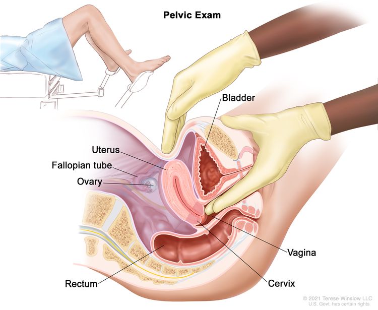 Pelvic exam. A doctor or nurse inserts one or two lubricated, gloved fingers of one hand into the vagina and presses on the lower abdomen with the other hand. This is done to feel the size, shape, and position of the uterus and ovaries. The vagina, cervix, fallopian tubes, and rectum are also checked.
 - CA 125assay: A test that measures the level of CA 125 in the blood. CA 125 is a substance released by cells into the bloodstream. An increased CA 125 level can be a sign of cancer or another condition such as endometriosis.
- Ultrasound exam: A procedure in which high-energy sound waves (ultrasound) are bounced off internal tissues or organs in the abdomen, and make echoes. The echoes form a picture of body tissues called a sonogram. The picture can be printed to be looked at later.
Some patients may have a transvaginal ultrasound.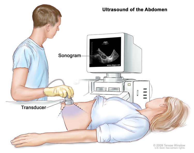 Abdominal ultrasound. An ultrasound transducer connected to a computer is passed over the surface of the abdomen. The ultrasound transducer bounces sound waves off internal organs and tissues to make echoes that form a sonogram (computer picture).
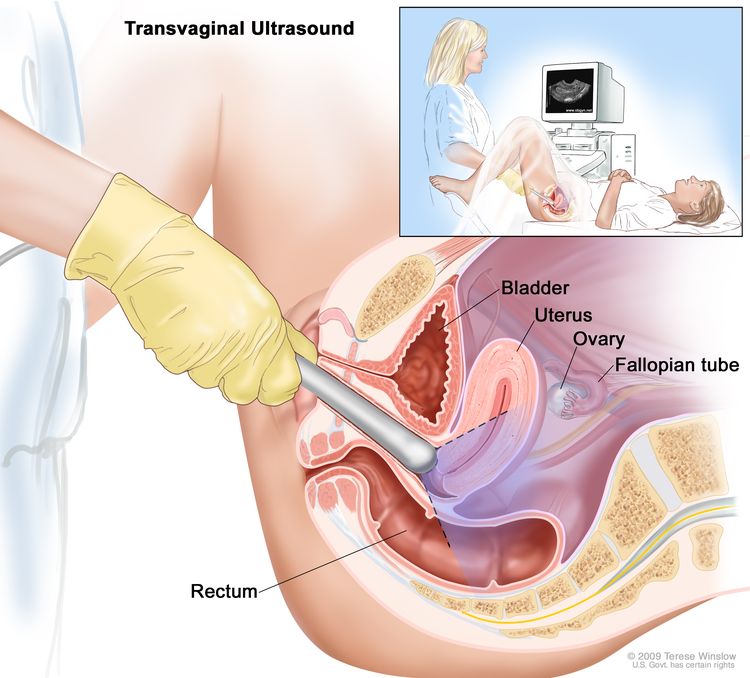 Transvaginal ultrasound. An ultrasound probe connected to a computer is inserted into the vagina and is gently moved to show different organs. The probe bounces sound waves off internal organs and tissues to make echoes that form a sonogram (computer picture).
 - CT scan (CAT scan): A procedure that makes a series of detailed pictures of areas inside the body, taken from different angles. The pictures are made by a computer linked to an x-ray machine. A dye may be injected into a vein or swallowed to help the organs or tissues show up more clearly. This procedure is also called computed tomography, computerized tomography, or computerized axial tomography.
- PET scan (positron emission tomography scan): A procedure to find malignant tumor cells in the body. A very small amount of radioactiveglucose (sugar) is injected into a vein. The PET scanner rotates around the body and makes a picture of where glucose is being used in the body. Malignant tumor cells show up brighter in the picture because they are more active and take up more glucose than normal cells do.
- MRI (magnetic resonance imaging): A procedure that uses a magnet, radio waves, and a computer to make a series of detailed pictures of areas inside the body. This procedure is also called nuclear magnetic resonance imaging (NMRI).
- Chest x-ray: An x-ray of the organs and bones inside the chest. An x-ray is a type of energy beam that can go through the body and onto film, making a picture of areas inside the body.
- Biopsy: The removal of cells or tissues so they can be viewed under a microscope by a pathologist to check for signs of cancer. The tissue is usually removed during surgery to remove the tumor.
Certain factors affect treatment options and prognosis (chance of recovery).
The prognosis and treatment options depend on the following:
- The type of ovarian cancer and how much cancer there is.
- The stage and grade of the cancer.
- Whether the patient has extra fluid in the abdomen that causes swelling.
- Whether all of the tumor can be removed by surgery.
- Whether there are changes in the BRCA1 or BRCA2genes.
- The patient’s age and general health.
- Whether the cancer has just been diagnosed or has recurred (come back).
Stages of Ovarian Epithelial, Fallopian Tube, and Primary Peritoneal Cancer
Key Points for this Section
- After ovarian, fallopian tube, or peritoneal cancer has been diagnosed, tests are done to find out if cancer cells have spread within the ovaries or to other parts of the body.
- There are three ways that cancer spreads in the body.
- Cancer may spread from where it began to other parts of the body.
- The following stages are used for ovarian epithelial, fallopian tube, and primary peritoneal cancer:
- Stage I
- Stage II
- Stage III
- Stage IV
- Ovarian epithelial, fallopian tube, and primary peritoneal cancers are grouped for treatment as early or advanced cancer.
- Ovarian epithelial, fallopian tube, and primary peritoneal cancers can recur (come back) after treatment.
After ovarian, fallopian tube, or peritoneal cancer has been diagnosed, tests are done to find out if cancer cells have spread within the ovaries or to other parts of the body.
The process used to find out whether cancer has spread within the organ or to other parts of the body is called staging. The information gathered from the staging process determines the stage of the disease. It is important to know the stage in order to plan treatment. The results of the tests used to diagnose cancer are often also used to stage the disease. (See the General Information section for tests and procedures used to diagnose and stage ovarian, fallopian tube, and peritoneal cancer.)
There are three ways that cancer spreads in the body.
Cancer can spread through tissue, the lymph system, and the blood:
- Tissue. The cancer spreads from where it began by growing into nearby areas.
- Lymph system. The cancer spreads from where it began by getting into the lymph system. The cancer travels through the lymph vessels to other parts of the body.
- Blood. The cancer spreads from where it began by getting into the blood. The cancer travels through the blood vessels to other parts of the body.
Cancer may spread from where it began to other parts of the body.
When cancer spreads to another part of the body, it is called metastasis. Cancer cells break away from where they began (the primary tumor) and travel through the lymph system or blood.
- Lymph system. The cancer gets into the lymph system, travels through the lymph vessels, and forms a tumor (metastatic tumor) in another part of the body.
- Blood. The cancer gets into the blood, travels through the blood vessels, and forms a tumor (metastatic tumor) in another part of the body.
The metastatic tumor is the same type of cancer as the primary tumor. For example, if ovarian epithelial cancer spreads to the lung, the cancer cells in the lung are actually ovarian epithelial cancer cells. The disease is metastatic ovarian epithelial cancer, not lung cancer.
metastasis: how cancer spreadsMany cancer deaths are caused when cancer moves from the original tumor and spreads to other tissues and organs. This is called metastatic cancer. This animation shows how cancer cells travel from the place in the body where they first formed to other parts of the body.The following stages are used for ovarian epithelial, fallopian tube, and primary peritoneal cancer:
Stage I
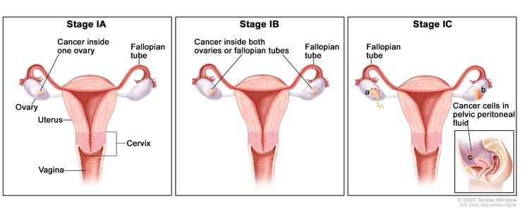 In stage IA, cancer is found inside a single ovary or fallopian tube. In stage IB, cancer is found inside both ovaries or fallopian tubes. In stage IC, cancer is found inside one or both ovaries or fallopian tubes and one of the following is true: (a) either the tumor or the capsule (outer covering) of the ovary has ruptured (broken open), or (b) cancer is also found on the surface of the ovary or fallopian tube, or (c) cancer cells are found in the pelvic peritoneal fluid.
In stage I, cancer is found in one or both ovaries or fallopian tubes. Stage I is divided into stage IA, stage IB, and stage IC.
- Stage IA: Cancer is found inside a single ovary or fallopian tube.
- Stage IB: Cancer is found inside both ovaries or fallopian tubes.
- Stage IC: Cancer is found inside one or both ovaries or fallopian tubes and one of
the following is true:
- the tumor ruptured (broke open) during surgery; or
- the capsule (outer covering) of the ovary ruptured before surgery, or there is cancer on the surface of the ovary or fallopian tube; or
- cancer cells are found in the fluid of the peritoneal cavity (the body cavity that contains most of the organs in the abdomen) or in washings of the peritoneum (tissue lining the peritoneal cavity).
Stage II
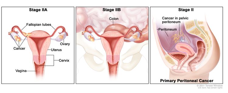 In stage IIA, cancer is found in one or both ovaries or fallopian tubes and has spread to the uterus and/or the fallopian tubes and/or the ovaries. In stage IIB, cancer is found in one or both ovaries or fallopian tubes and has spread to organs in the peritoneal cavity, such as the colon. In primary peritoneal cancer, cancer is found in the pelvic peritoneum and has not spread there from another part of the body.
In stage II, cancer is found in one or both ovaries or fallopian tubes and has spread into other areas of the pelvis, or primary peritoneal cancer is found within the pelvis. Stage II is divided into stage IIA and stage IIB.
- Stage IIA: Cancer has spread from where it first formed to the uterus and/or the fallopian tubes and/or the ovaries.
- Stage IIB: Cancer has spread from the ovary or fallopian tube to organs in the peritoneal cavity (the body cavity that contains most of the organs in the abdomen).
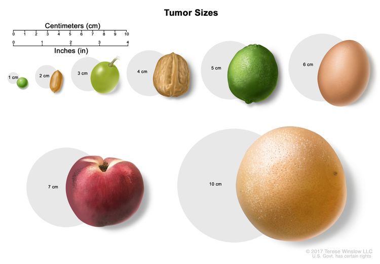 Tumor sizes are often measured in centimeters (cm) or inches. Common food items that can be used to show tumor size in cm include: a pea (1 cm), a peanut (2 cm), a grape (3 cm), a walnut (4 cm), a lime (5 cm or 2 inches), an egg (6 cm), a peach (7 cm), and a grapefruit (10 cm or 4 inches).
Stage III
In stage III, cancer is found in one or both ovaries or fallopian tubes, or is primary peritoneal cancer, and has spread outside the pelvis to other parts of the abdomen and/or to nearby lymph nodes. Stage III is divided into stage IIIA, stage IIIB, and stage IIIC.
- In stage IIIA, one of the following is true:
- Cancer has spread to lymph nodes behind the peritoneum only; or
- Cancer cells that can be seen only with a microscope have spread to the surface of the peritoneum outside the pelvis, such as the omentum (a fold of the peritoneum that surrounds the stomach and other organs in the abdomen). Cancer may have spread to nearby lymph nodes.
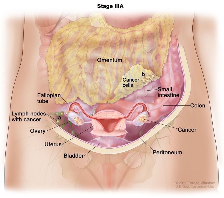 In stage IIIA, cancer is found in one or both ovaries or fallopian tubes and (a) cancer has spread to lymph nodes behind the peritoneum only, or (b) cancer cells that can be seen only with a microscope have spread to the surface of the peritoneum outside the pelvis, such as the omentum. Cancer may have also spread to nearby lymph nodes.
 - Stage IIIB: Cancer has spread to the peritoneum outside the pelvis, such as the omentum, and the cancer in the peritoneum is 2 centimeters or smaller. Cancer may have spread to lymph nodes behind the peritoneum.
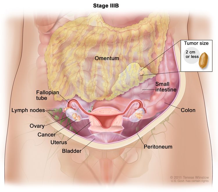 In stage IIIB, cancer is found in one or both ovaries or fallopian tubes and has spread to the peritoneum outside the pelvis, such as the omentum. The cancer in the omentum is 2 centimeters or smaller. Cancer may have also spread to lymph nodes behind the peritoneum.
 - Stage IIIC: Cancer has spread to the peritoneum outside the pelvis, such as the omentum, and the cancer in the peritoneum is
larger than 2 centimeters. Cancer may have spread to
lymph nodes behind the peritoneum or to the surface of the liver or spleen.
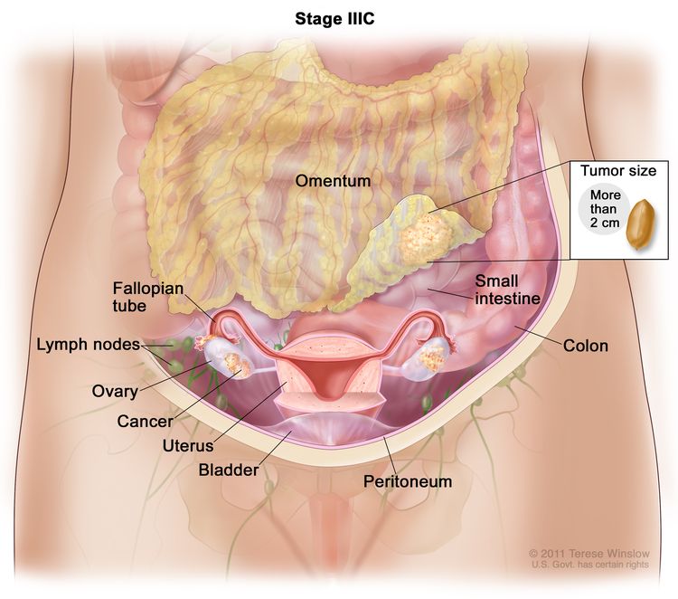 In stage IIIC, cancer is found in one or both ovaries or fallopian tubes and has spread to the peritoneum outside the pelvis, such as the omentum. The cancer in the omentum is larger than 2 centimeters. Cancer may have also spread to lymph nodes behind the peritoneum or to the surface of the liver or spleen (not shown).
Stage IV
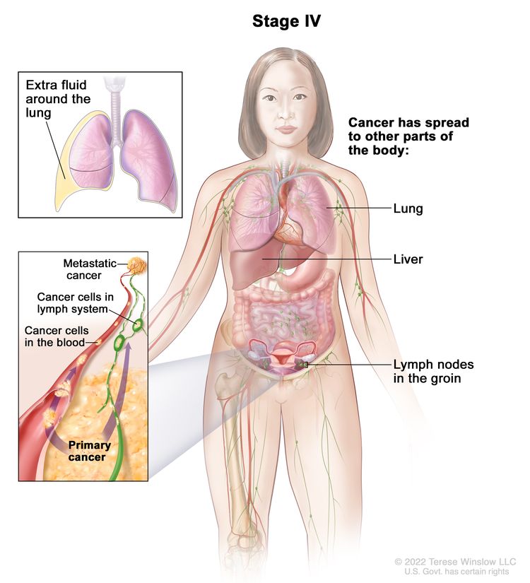 In stage IV, cancer has spread beyond the abdomen to other parts of the body. In stage IVA, cancer cells are found in extra fluid that builds up around the lungs. In stage IVB, cancer has spread to organs and tissues outside the abdomen, including the lung, liver, and lymph nodes in the groin.
In stage IV, cancer has spread beyond the abdomen to other parts of the body. Stage IV is divided into stage IVA and stage IVB.
- Stage IVA: Cancer cells are found in extra fluid that builds up around the lungs.
- Stage IVB: Cancer has spread to organs and tissues outside the abdomen, including lymph nodes in the groin.
Ovarian epithelial, fallopian tube, and primary peritoneal cancers are grouped for treatment as early or advanced cancer.
Stage I ovarian epithelial and fallopian tube cancers are treated as early cancers.
Stages II, III, and IV ovarian epithelial, fallopian tube, and primary peritoneal cancers are treated as advanced cancers.
Ovarian epithelial, fallopian tube, and primary peritoneal cancers can recur (come back) after treatment.
The cancer may come back in the same place or in other parts of the body. Persistent cancer is cancer that does not go away with treatment.
Treatment Option Overview
Key Points for this Section
- There are different types of treatment for patients with ovarian epithelial cancer.
- Three kinds of standard treatment are used.
- Surgery
- Chemotherapy
- Targeted therapy
- New types of treatment are being tested in clinical trials.
- Radiation therapy
- Immunotherapy
- Treatment for ovarian epithelial, fallopian tube, and primary peritoneal cancer may cause side effects.
- Patients may want to think about taking part in a clinical trial.
- Patients can enter clinical trials before, during, or after starting their cancer treatment.
- Follow-up tests may be needed.
There are different types of treatment for patients with ovarian epithelial cancer.
Different types of treatment are available for patients with ovarian epithelial cancer. Some treatments are standard, and some are being tested in clinical trials. A treatment clinical trial is a research study meant to help improve current treatments or obtain information on new treatments for patients with cancer. When clinical trials show that a new treatment is better than the treatment currently used as standard treatment, the new treatment may become the standard treatment. Patients with any stage of ovarian cancer may want to think about taking part in a clinical trial. Some clinical trials are open only to patients who have not started treatment.
Three kinds of standard treatment are used.
Surgery
Most patients have surgery to remove as much of the tumor as possible. Different types of surgery may include:
- Hysterectomy: Surgery to remove the uterus and, sometimes, the cervix. When only the uterus is removed, it is called a partial hysterectomy. When both the uterus and the cervix are removed, it is called a total hysterectomy. If the uterus and cervix are taken out through the vagina, the operation is called a vaginal hysterectomy. If the uterus and cervix are taken out through a large incision (cut) in the abdomen, the operation is called a total abdominal hysterectomy. If the uterus and cervix are taken out through a small incision (cut) in the abdomen using a laparoscope, the operation is called a total laparoscopic hysterectomy.
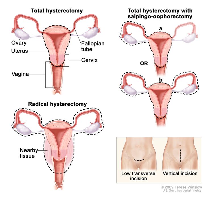 Hysterectomy. The uterus is surgically removed with or without other organs or tissues. In a total hysterectomy, the uterus and cervix are removed. In a total hysterectomy with salpingo-oophorectomy, (a) the uterus plus one (unilateral) ovary and fallopian tube are removed; or (b) the uterus plus both (bilateral) ovaries and fallopian tubes are removed. In a radical hysterectomy, the uterus, cervix, both ovaries, both fallopian tubes, and nearby tissue are removed. These procedures are done using a low transverse incision or a vertical incision.
 - Unilateral salpingo-oophorectomy: A surgical procedure to remove one ovary and one fallopian tube.
- Bilateral salpingo-oophorectomy: A surgical procedure to remove both ovaries and both fallopian tubes.
- Omentectomy: A surgical procedure to remove the omentum (tissue in the peritoneum that contains blood vessels, nerves, lymph vessels, and lymph nodes).
- Lymph node biopsy: The removal of all or part of a lymph node. A pathologist views the lymph node tissue under a microscope to check for cancer cells.
Chemotherapy
Chemotherapy is a cancer treatment that uses drugs to stop the growth of cancer cells, either by killing the cells or by stopping them from dividing. When chemotherapy is taken by mouth or injected into a vein or muscle, the drugs enter the bloodstream and can reach cancer cells throughout the body (systemic chemotherapy). When chemotherapy is placed directly into the cerebrospinal fluid, an organ, or a body cavity such as the abdomen, the drugs mainly affect cancer cells in those areas (regional chemotherapy).
A type of regional chemotherapy used to treat ovarian cancer is intraperitoneal (IP) chemotherapy. In IP chemotherapy, the anticancer drugs are carried directly into the peritoneal cavity (the space that contains the abdominal organs) through a thin tube.
Hyperthermic intraperitoneal chemotherapy (HIPEC) is a treatment used during surgery that is being studied for ovarian cancer. After the surgeon has removed as much tumor tissue as possible, warmed chemotherapy is sent directly into the peritoneal cavity.
Treatment with more than one anticancer drug is called combination chemotherapy.
The way the chemotherapy is given depends on the type and stage of the cancer being treated.
See Drugs Approved for Ovarian, Fallopian Tube, or Primary Peritoneal Cancer.
Targeted therapy
Targeted therapy is a type of treatment that uses drugs or other substances to identify and attack specific cancer cells.
Monoclonal antibodies are immune systemproteins made in the laboratory to treat many diseases, including cancer. As a cancer treatment, these antibodies can attach to a specific target on cancer cells or other cells that may help cancer cells grow. The antibodies are able to then kill the cancer cells, block their growth, or keep them from spreading. Monoclonal antibodies are given by infusion. They may be used alone or to carry drugs, toxins, or radioactive material directly to cancer cells. Monoclonal antibodies may be used in combination with chemotherapy as adjuvant therapy.
Bevacizumab is a monoclonal antibody and angiogenesis inhibitor that may be used with chemotherapy to treat ovarian epithelial cancer, fallopian tube cancer, or primary peritoneal cancer that has recurred (come back). It binds to a protein called vascular endothelial growth factor (VEGF) and may prevent the growth of new blood vessels that tumors need to grow. Other angiogenesis inhibitors are being studied in the treatment of advanced or recurrent ovarian cancer.
monoclonal antibodies: how monoclonal antibodies treat cancerHow do monoclonal antibodies work to treat cancer? This video shows how monoclonal antibodies, such as trastuzumab, pembrolizumab, and rituximab, block molecules cancer cells need to grow, flag cancer cells for destruction by the body’s immune system, or deliver harmful substances to cancer cells.Poly (ADP-ribose) polymerase inhibitors (PARP inhibitors) are targeted therapy drugs that block DNA repair and may cause cancer cells to die. Olaparib, rucaparib, and niraparib are PARP inhibitors that may be used as maintenance therapy to treat certain types of ovarian epithelial cancer, fallopian tube cancer, or primary peritoneal cancer that have recurred. Veliparib is a PARP inhibitor that is being studied in combination with chemotherapy to treat advanced ovarian cancer.
See Drugs Approved for Ovarian, Fallopian Tube, or Primary Peritoneal Cancer.
New types of treatment are being tested in clinical trials.
This summary section describes treatments that are being studied in clinical trials. It may not mention every new treatment being studied. Information about clinical trials is available from the NCI website.
Radiation therapy
Radiation therapy is a cancer treatment that uses high-energy x-rays or other types of radiation to kill cancer cells or keep them from growing. Some women receive a treatment called intraperitoneal radiation therapy, in which radioactive liquid is put directly in the abdomen through a catheter. Intraperitoneal radiation therapy is being studied to treat advanced ovarian cancer.
Immunotherapy
Immunotherapy is a treatment that uses the patient’s immune system to fight cancer. Substances made by the body or made in a laboratory are used to boost, direct, or restore the body’s natural defenses against cancer. This cancer treatment is a type of biologic therapy.
Vaccine therapy is a cancer treatment that uses a substance or group of substances to stimulate the immune system to find the tumor and kill it. Vaccine therapy is being studied to treat advanced ovarian cancer.
Treatment for ovarian epithelial, fallopian tube, and primary peritoneal cancer may cause side effects.
For information about side effects caused by treatment for cancer, see our Side Effects page.
Patients may want to think about taking part in a clinical trial.
For some patients, taking part in a clinical trial may be the best treatment choice. Clinical trials are part of the cancer research process. Clinical trials are done to find out if new cancer treatments are safe and effective or better than the standard treatment.
Many of today's standard treatments for cancer are based on earlier clinical trials. Patients who take part in a clinical trial may receive the standard treatment or be among the first to receive a new treatment.
Patients who take part in clinical trials also help improve the way cancer will be treated in the future. Even when clinical trials do not lead to effective new treatments, they often answer important questions and help move research forward.
Patients can enter clinical trials before, during, or after starting their cancer treatment.
Some clinical trials only include patients who have not yet received treatment. Other trials test treatments for patients whose cancer has not gotten better. There are also clinical trials that test new ways to stop cancer from recurring (coming back) or reduce the side effects of cancer treatment.
Clinical trials are taking place in many parts of the country. Information about clinical trials supported by NCI can be found on NCI’s clinical trials search webpage. Clinical trials supported by other organizations can be found on the ClinicalTrials.gov website.
Follow-up tests may be needed.
Some of the tests that were done to diagnose the cancer or to find out the stage of the cancer may be repeated. Some tests will be repeated in order to see how well the treatment is working. Decisions about whether to continue, change, or stop treatment may be based on the results of these tests.
Some of the tests will continue to be done from time to time after treatment has ended. The results of these tests can show if your condition has changed or if the cancer has recurred (come back). These tests are sometimes called follow-up tests or check-ups.
stage I ovarian epithelial cancerfallopian tube cancerTreatment of Early Ovarian Epithelial and Fallopian Tube Cancer
For information about the treatments listed below, see the Treatment Option Overview section.
Treatment of early ovarian epithelial cancer or fallopian tube cancer may include the following:
- Hysterectomy, bilateral salpingo-oophorectomy, and omentectomy. Lymph nodes and other tissues in the pelvis and abdomen are removed and checked under a microscope for cancercells. Chemotherapy may be given after surgery.
- Unilateral salpingo-oophorectomy may be done in certain women who wish to have children. Chemotherapy may be given after surgery.
Use our clinical trial search to find NCI-supported cancer clinical trials that are accepting patients. You can search for trials based on the type of cancer, the age of the patient, and where the trials are being done. General information about clinical trials is also available.
stage II ovarian epithelial cancerstage III ovarian epithelial cancerstage IV ovarian epithelial cancerfallopian tube cancerprimary peritoneal cavity cancerTreatment of Advanced Ovarian Epithelial, Fallopian Tube, and Primary Peritoneal Cancer
For information about the treatments listed below, see the Treatment Option Overview section.
Treatment of advanced ovarian epithelial cancer, fallopian tube cancer, or primary peritoneal cancer may include the following:
- Hysterectomy, bilateral salpingo-oophorectomy, and omentectomy. Lymph nodes and other tissues in the pelvis and abdomen are removed and checked under a microscope to look for cancercells. Surgery is followed by one of the following:
- Intravenous chemotherapy.
- Intraperitoneal chemotherapy.
- Chemotherapy and targeted therapy (bevacizumab).
- Chemotherapy and targeted therapy with a poly (ADP-ribose) polymerase (PARP) inhibitor.
- Chemotherapy and targeted therapy followed by surgery (possibly followed by intraperitoneal chemotherapy).
- Chemotherapy alone for patients who cannot have surgery.
- Targeted therapy with a PARP inhibitor (olaparib, rucaparib, niraparib).
- A clinical trial of targeted therapy with a PARP inhibitor (veliparib).
Use our clinical trial search to find NCI-supported cancer clinical trials that are accepting patients. You can search for trials based on the type of cancer, the age of the patient, and where the trials are being done. General information about clinical trials is also available.
recurrent ovarian epithelial cancerfallopian tube cancerprimary peritoneal cavity cancerTreatment of Recurrent or Persistent Ovarian Epithelial, Fallopian Tube, and Primary Peritoneal Cancer
For information about the treatments listed below, see the Treatment Option Overview section.
Treatment of recurrentovarian epithelial cancer, fallopian tube cancer, or primary peritoneal cancer may include the following:
- Chemotherapy using one or more anticancer drugs.
- Targeted therapy with a poly (ADP-ribose) polymerase (PARP) inhibitor (olaparib, rucaparib, or niraparib) with or without chemotherapy.
- Chemotherapy and/or targeted therapy (bevacizumab).
- A clinical trial of hyperthermicintraperitoneal chemotherapy (HIPEC) during surgery.
- A clinical trial of a new treatment.
Use our clinical trial search to find NCI-supported cancer clinical trials that are accepting patients. You can search for trials based on the type of cancer, the age of the patient, and where the trials are being done. General information about clinical trials is also available.
To Learn More About Ovarian Epithelial, Fallopian Tube, and Primary Peritoneal Cancer
For more information from the National Cancer Institute about ovarian epithelial, fallopian tube, and primary peritoneal cancer, see the following:
- Ovarian, Fallopian Tube, and Primary Peritoneal Cancer Home Page
- Ovarian, Fallopian Tube, and Primary Peritoneal Cancer Prevention
- Ovarian, Fallopian Tube, and Primary Peritoneal Cancer Screening
- Childhood Ovarian Cancer Treatment
- Drugs Approved for Ovarian, Fallopian Tube, or Primary Peritoneal Cancer
- Targeted Cancer Therapies
- BRCA Gene Mutations: Cancer Risk and Genetic Testing
- Genetic Testing for Inherited Cancer Susceptibility Syndromes
For general cancer information and other resources from the National Cancer Institute, see the following:
- About Cancer
- Staging
- Chemotherapy and You: Support for People With Cancer
- Radiation Therapy and You: Support for People With Cancer
- Coping with Cancer
- Questions to Ask Your Doctor about Cancer
- For Survivors and Caregivers
About This PDQ Summary
About PDQ
Physician Data Query (PDQ) is the National Cancer Institute's (NCI's) comprehensive cancer information database. The PDQ database contains summaries of the latest published information on cancer prevention, detection, genetics, treatment, supportive care, and complementary and alternative medicine. Most summaries come in two versions. The health professional versions have detailed information written in technical language. The patient versions are written in easy-to-understand, nontechnical language. Both versions have cancer information that is accurate and up to date and most versions are also available in Spanish.
PDQ is a service of the NCI. The NCI is part of the National Institutes of Health (NIH). NIH is the federal government’s center of biomedical research. The PDQ summaries are based on an independent review of the medical literature. They are not policy statements of the NCI or the NIH.
Purpose of This Summary
This PDQ cancer information summary has current information about the treatment of ovarian epithelial, fallopian tube, and primary peritoneal cancer. It is meant to inform and help patients, families, and caregivers. It does not give formal guidelines or recommendations for making decisions about health care.
Reviewers and Updates
Editorial Boards write the PDQ cancer information summaries and keep them up to date. These Boards are made up of experts in cancer treatment and other specialties related to cancer. The summaries are reviewed regularly and changes are made when there is new information. The date on each summary ("Updated") is the date of the most recent change.
The information in this patient summary was taken from the health professional version, which is reviewed regularly and updated as needed, by the PDQ Adult Treatment Editorial Board.
Clinical Trial Information
A clinical trial is a study to answer a scientific question, such as whether one treatment is better than another. Trials are based on past studies and what has been learned in the laboratory. Each trial answers certain scientific questions in order to find new and better ways to help cancer patients. During treatment clinical trials, information is collected about the effects of a new treatment and how well it works. If a clinical trial shows that a new treatment is better than one currently being used, the new treatment may become "standard." Patients may want to think about taking part in a clinical trial. Some clinical trials are open only to patients who have not started treatment.
Clinical trials can be found online at NCI's website. For more information, call the Cancer Information Service (CIS), NCI's contact center, at 1-800-4-CANCER (1-800-422-6237).
Permission to Use This Summary
PDQ is a registered trademark. The content of PDQ documents can be used freely as text. It cannot be identified as an NCI PDQ cancer information summary unless the whole summary is shown and it is updated regularly. However, a user would be allowed to write a sentence such as “NCI’s PDQ cancer information summary about breast cancer prevention states the risks in the following way: [include excerpt from the summary].”
The best way to cite this PDQ summary is:
PDQ® Adult Treatment Editorial Board. PDQ Ovarian Epithelial, Fallopian Tube, and Primary Peritoneal Cancer Treatment. Bethesda, MD: National Cancer Institute. Updated
Images in this summary are used with permission of the author(s), artist, and/or publisher for use in the PDQ summaries only. If you want to use an image from a PDQ summary and you are not using the whole summary, you must get permission from the owner. It cannot be given by the National Cancer Institute. Information about using the images in this summary, along with many other images related to cancer can be found in Visuals Online. Visuals Online is a collection of more than 3,000 scientific images.
Disclaimer
The information in these summaries should not be used to make decisions about insurance reimbursement. More information on insurance coverage is available on Cancer.gov on the Managing Cancer Care page.
Contact Us
More information about contacting us or receiving help with the Cancer.gov website can be found on our Contact Us for Help page. Questions can also be submitted to Cancer.gov through the website’s E-mail Us.