National Cancer Institute
Post Date: Oct 30, 2019
Cardiopulmonary syndromes are conditions of the heart and lung and can occur in some cancers. They include shortness of breath (dyspnea), chronic cough, pleural and pericardial effusion, and superior vena cava syndrome. Learn more about these conditions in this expert-reviewed summary.
Cardiopulmonary Syndromes
Cardiopulmonary Syndromes Overview
Cardiopulmonarysyndromes are conditions of the heart and lung that may be caused by cancer or by other health problems. Five cardiopulmonary syndromes that may be caused by cancer are covered in this summary:
- Dyspnea (shortness of breath).
- Chronic cough.
- Malignant pleural effusion (extra fluid around the lungs caused by cancer).
- Malignant pericardial effusion (extra fluid in the sac around the heart caused by cancer).
- Superior vena cava syndrome (a blocked superior vena cava, the large vein that takes blood back to the heart).
This summary is about cardiopulmonary syndromes in adults and children with cancer. Section titles show when the information is about children.
Dyspnea During Advanced Cancer
Key Points for this Section
- Many conditions can cause dyspnea.
- A physical exam and health history are important in finding out the cause of dyspnea.
- There are different ways to treat the causes of dyspnea in cancer patients.
- Treatment of dyspnea will depend on what is causing it.
- Treatment may be to control the signs and symptoms of dyspnea.
Many conditions can cause dyspnea.
Dyspnea is the feeling that you can't catch your breath or you can't breathe in enough air. It also may be called shortness of breath, breathlessness, or air hunger. In cancer patients, dyspnea can be caused by the following:
- Tumor-related effects:
- The tumor blocks the airways in the chest and lung or it blocks the vein that carries blood through the chest to the heart.
- The tumor causes extra fluid to build up in the space between the thin layer of tissue covering the lung and the thin layer of tissue covering the chest wall (pleural effusion), between the sac that covers the heart and the heart (pericardial effusion), or in the abdominalcavity (ascites).
- Superior vena cava syndrome. (a group of signs or symptoms that occur when the superior vena cava is partly blocked).
- Carcinomatous lymphangitis (inflammation of the lymph vessels).
- Pneumonia and other chest infections.
- Pneumonitis (inflammation of the lungs).
- Blood clots or tumor cells break loose and block a blood vessel in the lungs.
- Paralysis of part of the diaphragm (a muscle used for breathing).
- Breathing muscles get weaker.
- Treatment-related effects:
- Damage to the lung caused by radiation therapy or chemotherapy. A small number of women who receive radiation therapy for breast cancer develop postradiation bronchiolitis obliterans, a condition in which the bronchioles (tiny branches of air tubes in the lungs) become inflamed and blocked.
- Weakened heart muscle caused by chemotherapy.
- Immunotherapy-related pneumonitis. Pneumonitis linked to immunotherapy with a checkpoint inhibitor is uncommon but can be serious or life-threatening. In one study of patients treated for immunotherapy-related pneumonitis, melanoma and non-small cell lung cancer were the most common cancers. The length of time that patients received immunotherapy before developing pneumonitis varied from several days to over a year. In addition, pneumonitis seemed to occur earlier in patients who received combination therapy than in those who received one type of treatment.
- Conditions that are not related to cancer:
- Chronic obstructive pulmonary disease (COPD), such as chronicbronchitis or emphysema.
- Bronchospasm. The muscles in the airways contract and cause spasms.
- Congestive heart failure.
- Anemia.
- Conditions with no known physical cause, such as anxiety.
A physical exam and health history are important in finding out the cause of dyspnea.
Diagnostic tests and procedures include the following:
- Physical exam and health history: An exam of the body to check general signs of health, including checking for signs or symptoms of dyspnea, such as breathing fast or using the neck or chest muscles to breathe. A history of your health habits and past illnesses and treatments will also be taken. Your doctor will also ask about when the dyspnea occurs, what it feels like, other signs or symptoms that happen at the same time as the dyspnea, and anything that makes it better or worse.
- Functional assessment: An exam to check how the dyspnea affects your ability to perform activities of daily living such as eating, bathing, or climbing stairs. This exam may include a 6-minute walk test (6MWT) to measure how far you can walk on a flat, hard surface in 6 minutes.
- Chest x-ray:
An x-ray of the organs and bones inside the chest. An x-ray is a type of energy beam that can go through the body and onto film, making a picture of areas inside the body.
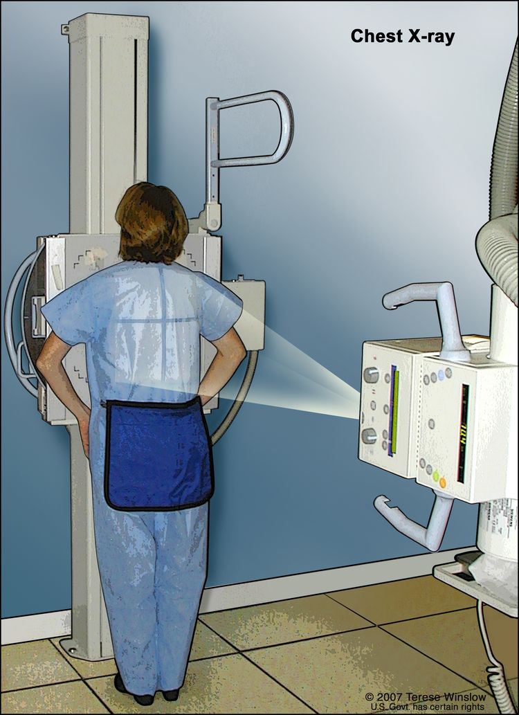 A chest x-ray is used to take pictures of the structures and organs inside the chest. X-rays pass through the patient's body onto film or a computer.
 - CT scan (CAT scan): A procedure that makes a series of detailed pictures of areas inside the body, taken from different angles. The pictures are made by a computer linked to an x-ray machine. A dye may be injected into a vein or swallowed to help the organs or tissues show up more clearly. This procedure is also called computed tomography, computerized tomography, or computerized axial tomography.
- Complete blood count: A procedure in which a sample of blood is taken and checked for the following:
- The number of red blood cells, white blood cells, and platelets.
- The amount of hemoglobin (the protein that carries oxygen) in the red blood cells.
- The portion of the blood sample made of red blood cells.
- Oxygen saturation test: A procedure to check for the amount of oxygen being carried by the red blood cells. A lower than normal amount of oxygen may be a sign of lung disease or other health problems. One method uses a device clipped to the finger. The device senses the amount of oxygen in the blood flowing through the small blood vessels in the finger. Another method uses a sample of blood taken from an artery, usually in the wrist, that is tested for the amount of oxygen.
- Maximum inspiratory pressure (MIP) test: The MIP is the highest pressure that can be reached in the lungs when you take a deep breath. When you breathe through a device called a manometer, the device measures the pressure. The information is sent to a computer. The pressure level shows how strong the breathing muscles are.
There are different ways to treat the causes of dyspnea in cancer patients.
Treatment may include the following:
- Radiation therapy: Radiation therapy is a cancer treatment that uses high-energy x-rays or other types of radiation to kill cancer cells or keep them from growing. External radiation therapy uses a machine outside the body to send radiation toward the cancer.
- Chemotherapy: Chemotherapy is a cancer treatment that uses drugs to stop the growth of cancer cells, either by killing the cells or by stopping them from dividing. When chemotherapy is taken by mouth or injected into a vein or muscle, the drugs enter the bloodstream and can reach cancer cells throughout the body (systemic chemotherapy). When chemotherapy is placed directly into the cerebrospinal fluid, an organ, or a body cavity such as the abdomen, the drugs mainly affect cancer cells in those areas (regional chemotherapy). The way the chemotherapy is given depends on the type and stage of the cancer being treated.
- Laser therapy for tumors inside large airways: Use of a laser beam (a narrow beam of intense light) as a knife to remove the tumor.
- Cauterization of tumors inside large airways: Use of a hot instrument, an electric current, or a caustic substance to destroy the tumor.
- Procedures to remove fluid that has built up around the lungs (malignant pleural effusion), around the heart (malignant pericardial effusion ), or in the abdominal cavity (ascites). (See the sections on controlling the signs and symptoms of malignant pleural effusion and malignant pericardial effusion for more information.)
- Stent placement: Surgery to place a stent (thin tube) in an airway to keep it open. This may be done if a large airway is blocked by a tumor that is pressing on it from the outside.
- Medicine:
- Steroid drugs for inflamed or swollen lymph vessels in the lungs.
- Antibiotics for chest infections. These may be used along with breathing treatments.
- Anticoagulants for blood clots that are blocking blood vessels in the lungs.
- Bronchodilators that are inhaled to open up the bronchioles (small airways) in the lungs.
- Diuretics and other drugs for heart failure.
- Blood transfusions for anemia.
Treatment of dyspnea will depend on what is causing it.
The treatment of dyspnea depends on its cause, as follows:
| If the dyspnea is caused by: | Then the treatment may be: |
|---|---|
| Tumor blocking the large or small airways in the chest or lung | • Radiation therapy. |
| • Chemotherapy, for tumors that usually respond quickly to this treatment. | |
| • Laser surgery to remove the tumor. | |
| • Cauterization of tumors. | |
| • Stent placement to keep airway open. | |
| Pleural effusion | • Removal of the extra fluid around the lung using a needle or chest drain. |
| Pericardial effusion | • Removal of the extra fluid around the heart using a needle. |
| • Intrapericardial chemotherapy. | |
| • Surgery. | |
| Ascites | • Removal of the extra fluid in the abdominal cavity using a needle. |
| Carcinomatous lymphangitis | • Steroid therapy. |
| • Chemotherapy, for tumors that usually respond quickly to this treatment. | |
| Superior vena cava syndrome | • Chemotherapy, for tumors that usually respond quickly to this treatment. |
| • Radiation therapy. | |
| • Surgery to place a stent in the superior vena cava to keep it open. | |
| • Opioids and/or steroid therapy. | |
| Chest infections | • Antibiotics. |
| • Breathing treatments. | |
| Pulmonary embolism | • Anticoagulants. |
| Bronchospasms or chronic obstructive pulmonary disease | • Bronchodilators. |
| • Inhaled steroids. | |
| Postradiation bronchiolitis obliterans | • Steroid therapy. |
| Heart failure | • Diuretics and other heart medicines. |
| Anemia | • Blood transfusion |
| Checkpoint inhibitor immunotherapy–related pneumonitis | • Withholding drug therapy. |
| • Corticosteroids. | |
| • Close follow-up. |
Treatment may be to control the signs and symptoms of dyspnea.
Treatment to control the signs and symptoms of dyspnea may include the following:
- Oxygen therapy: If you cannot breathe in enough oxygen, you may be given extra oxygen to inhale from a tank. Devices that deliver a high flow of oxygen or air mixed with oxygen may also be prescribed.
- Medicines: Opioids, such as morphine, may help with distress, fatigue, and the feeling that you cannot get enough air. Other drugs may be used to treat dyspnea that is related to panic disorder or severe anxiety.
- Non-drug treatments:
- Breathing methods, such as breathing with the lips pursed (almost closed).
- Using a fan to blow cold air across the cheek.
- Meditation.
- Relaxation training.
- Biofeedback.
- Talk therapy to relieve anxiety.
Chronic Coughing
Key Points for this Section
- Chronic coughing may cause much physical distress.
- It may be possible to treat the cause of chronic coughing.
- Medicines may be used to control chronic coughing.
Chronic coughing may cause much physical distress.
Chronic cough may cause pain, trouble sleeping, dyspnea, or fatigue. The causes of chronic coughing are almost the same as the causes of dyspnea. See Dyspnea section for list of causes.
It may be possible to treat the cause of chronic coughing.
Treatments may include:
- Radiation therapy for a tumor that is blocking the airway.
- Placement of a stent (tube) to close a fistula caused by esophageal cancer.
- Draining the fluid of a pleural effusion.
- Corticosteroids for lymphangitic carcinomatosis.
Medicines may be used to control chronic coughing.
Medicines may include:
- Medicines to stop the cough, including opioids.
- Medicine that breaks down mucus.
- An inhaleddrug for chronic coughing related to lung cancer.
Malignant Pleural Effusion
Key Points for this Section
- Pleural effusion is extra fluid around the lungs.
- Pleural effusion may be caused by cancer, cancer treatment, or other conditions.
- Signs and symptoms of pleural effusion include dyspnea (shortness of breath) and cough.
- Finding out the cause of pleural effusion will help plan the treatment.
- Treatment may be to control signs and symptoms of pleural effusion and improve quality of life.
Pleural effusion is extra fluid around the lungs.
The pleural cavity is the space between the pleura (thin layer of tissue) that covers the outer surface of each lung and lines the inner wall of the chest cavity. Pleural tissue usually makes a small amount of fluid that helps the lungs move smoothly in the chest while a person is breathing. A pleural effusion is extra fluid in the pleural cavity. The fluid presses on the lungs and makes it hard to breathe.
Pleural effusion may be caused by cancer, cancer treatment, or other conditions.
A pleural effusion may be malignant (caused by cancer) or nonmalignant (caused by a condition that is not cancer). Malignant pleural effusion is a common problem for patients who have certain cancers. Lung cancer, breast cancer, lymphoma, and leukemia cause most malignant effusions.
Pleural effusion also may be caused by radiation therapy, chemotherapy, a collapsed lung, or cancer that has spread to lymph nodes. Some cancer patients have conditions such as congestive heart failure, pneumonia, blood clot in the lung, or poor nutrition that may lead to a pleural effusion.
Signs and symptoms of pleural effusion include dyspnea (shortness of breath) and cough.
These and other signs and symptoms may be caused by a pleural effusion. Talk to your doctor if you have any of the following problems:
- Dyspnea (shortness of breath).
- Cough.
- An uncomfortable feeling or pain in the chest.
Finding out the cause of pleural effusion will help plan the treatment.
Treatment for a malignant pleural effusion is different from treatment for a nonmalignant effusion, so the right diagnosis is important. Diagnostic tests used to find the cause of the pleural effusion include the following:
- Chest x-ray: An x-ray of the organs and bones inside the chest. An x-ray is a type of energy beam that can go through the body and onto film, making a picture of areas inside the body.A chest x-ray is used to take pictures of the structures and organs inside the chest. X-rays pass through the patient's body onto film or a computer.
- CT scan: A procedure that makes a series of detailed pictures of areas inside the body, taken from different angles. The pictures are made by a computer linked to an x-ray machine. A dye may be injected into a vein or swallowed to help the organs or tissues show up more clearly. This procedure is also called computed tomography, computerized tomography, or computerized axial tomography.
- Ultrasound exam: A procedure in which high-energy sound waves (ultrasound) are bounced off internal tissues or organs and make echoes. The echoes form a picture of body tissues called a sonogram.
- Thoracentesis: The removal of fluid from the space between the lining of the chest and the lung, using a needle. A pathologist views the fluid under a microscope to look for cancer cells. This procedure may be used to reduce pressure on the lungs.
- Biopsy: The removal of cells or tissues so they can be viewed under a microscope by a pathologist to check for signs of cancer. If thoracentesis is not possible, a biopsy may be done during a thoracoscopy. A thoracoscopy is a procedure to look at the organs inside the chest to check for abnormal areas. An incision (cut) is made between two ribs and a thoracoscope (a thin, lighted tube with a lens for viewing) is inserted into the chest. A cutting tool at the end of the thoracoscope is used to remove a sample of tissue.
- Flow cytometry: A laboratory test that measures the number of cells in a sample, the percentage of live cells in a sample, and certain characteristics of the cells, such as size, shape, and the presence of tumor (or other) markers on the cell surface. The cells from a sample of a patient’s blood, bone marrow, or other tissue are stained with a fluorescent dye, placed in a fluid, and then passed one at a time through a beam of light. The test results are based on how the cells that were stained with the fluorescent dye react to the beam of light.
The type of cancer, previous treatment for cancer, and your choices also are important in planning treatment.
Treatment may be to control signs and symptoms of pleural effusion and improve quality of life.
A malignant pleural effusion often occurs in cancer that is advanced, cannot be removed by surgery, or continues to grow or spread during treatment. It is also common during the last few weeks of life. The goal of treatment is usually palliative, to relieve signs and symptoms and improve quality of life.
Treatment of the signs and symptoms of malignant pleural effusion includes the following:
- Thoracentesis. Thoracentesis is a procedure to remove extra fluid from the pleural cavity between the lung and the chest wall using a needle. Removal of the fluid may help to relieve severe symptoms for a short time. A few days after the extra fluid is removed, it is likely it will begin to come back. The risk of a thoracentesis includes bleeding, infection, collapsed lung, fluid in the lungs, and a sudden drop in blood pressure.
- Indwelling pleural catheter (IPC). An indwelling pleural catheter (IPC) is a small tube that is inserted and left in place to keep fluid from building up around the lungs. One end of the tube stays inside the chest and the other passes outside the body to allow fluid to drain. This type of catheter may be used for long-term care so that a separate procedure won't need to be done each time draining is needed. Risks of IPCs include infection and blockage of the catheter.
- Pleurodesis. This is a procedure to close the pleural space so that fluid cannot collect there. Fluid is first removed by thoracentesis, using a chest tube. A drug that causes the pleural space to close is then inserted into the space through a chest tube. Drugs such as bleomycin or talc may be used.
- Surgery. Surgery may be done to put in a shunt (tube) to carry the fluid from the pleural cavity to the abdominal cavity, where the fluid is easier to remove. Pleurectomy is another type of surgery that may be used. In this procedure, the part of the pleura that lines the chest cavity is removed.
Malignant Pericardial Effusion
Key Points for this Section
- Pericardial effusion is extra fluid around the heart.
- Pericardial effusion may be caused by cancer or other conditions.
- Signs and symptoms of pericardial effusion include dyspnea (shortness of breath) and cough.
- Pericardial effusion usually occurs in advanced cancer.
- Treatment may be to control the symptoms of pericardial effusion and improve quality of life.
Pericardial effusion is extra fluid around the heart.
Pericardial effusion is extra fluid inside the sac that surrounds the heart. The extra fluid causes pressure on the heart, which stops it from pumping blood normally. If fluid builds up, a condition called cardiac tamponade may occur. In cardiac tamponade, the heart cannot pump enough blood to the rest of the body. This is life-threatening and must be treated right away.
Pericardial effusion may be caused by cancer or other conditions.
A pericardial effusion may be malignant (caused by cancer) or nonmalignant (caused by a condition that is not cancer). Malignant pericardial effusion is common in lung cancer, breast cancer, melanoma, lymphoma, and leukemia patients. Pericarditis (swelling of tissues around the heart), a heart attack, hypothyroidism, or systemic lupus erythematosus are examples of nonmalignant causes of pericardial effusion. Radiation therapy or chemotherapy may cause pericarditis, leading to pericardial effusion.
Signs and symptoms of pericardial effusion include dyspnea (shortness of breath) and cough.
At first, a pericardial effusion may not cause any signs or symptoms. These and other signs and symptoms may be caused by a pericardial effusion or cardiac tamponade. Check with your doctor if you have any of the following:
- Dyspnea (shortness of breath).
- Cough.
- Chest pain or pressure.
- Trouble breathing while lying flat.
- Fast heart beat or breathing.
- Feeling faint.
- Pressure in the upper abdomen.
- Tiredness.
- Weakness.
Pericardial effusion usually occurs in advanced cancer.
Pericardial effusion usually occurs in advanced cancer or in the last few weeks of life. During these times, it may be more important to relieve the symptoms than to diagnose the condition. However, in some cases, the following tests and procedures may be used to diagnose pericardial effusion:
- Chest x-ray: An x-ray of the organs and bones inside the chest. An x-ray is a type of energy beam that can go through the body and onto film, making a picture of areas inside the body.A chest x-ray is used to take pictures of the structures and organs inside the chest. X-rays pass through the patient's body onto film or a computer.
- Echocardiography: A procedure in which high-energy sound waves (ultrasound) are bounced off internal tissues or organs of the chest. The echoes form a picture of the heart's position, motion of the walls, and internal parts such as the valves.
- Electrocardiogram (EKG or ECG): A line graph recording of the heart's electrical activity to check its rate and rhythm. A number of electrodes (small pads) are placed on the patient’s chest, arms, and legs. The electrodes are connected by wires to the EKG machine. Heart activity is then recorded on paper. Electrical activity that is faster or slower than normal may be a sign of heart problems.
- Pericardiocentesis: A procedure to remove fluid from the pericardium using a needle inserted through the chest wall. The doctor may use echocardiography to watch the movement of the heart and needle inside the chest. The fluid is viewed under a microscope by a pathologist to check for cancer cells or signs of infection. This procedure can also be used to treat pericardial effusion. Removing the fluid lessens the pressure on the heart.
- Flow cytometry: A laboratory test that measures the number of cells in a sample, the percentage of live cells in a sample, and certain characteristics of the cells, such as size, shape, and the presence of tumor (or other) markers on the cell surface. The cells from a sample of a patient’s blood, bone marrow, or other tissue are stained with a fluorescent dye, placed in a fluid, and then passed one at a time through a beam of light. The test results are based on how the cells that were stained with the fluorescent dye react to the beam of light.
Treatment may be to control the symptoms of pericardial effusion and improve quality of life.
The goal of treatment is usually palliative, to relieve symptoms and improve quality of life. If a malignant pericardial effusion is severe, it is usually controlled by draining the fluid.
Treatment options include the following:
- Pericardiocentesis. A procedure to remove the extra fluid from the sac around the heart using a needle inserted through the chest wall. The doctor may use echocardiography to watch the movement of the heart and needle inside the chest. Removing the fluid can reduce pressure on the heart. In some patients, fluid may again collect in the sac around the heart after pericardiocentesis. A catheter (flexible tube used to put fluids into or take blood out of a vein) may be inserted and left in place for several days until there is little to no fluid drainage. For patients with advanced cancer, this procedure may be used instead of more serious surgery.
- Pericardial sclerosis. A procedure to close the pericardial space so fluid cannot collect in the sac around the heart. Fluid is first removed by pericardiocentesis. A drug or chemical is then injected through a catheter (flexible tube used to put fluids into or take blood out of a vein) into the pericardial space to cause it to close. Three or more treatments may be needed to completely close the pericardial space.
- Pericardiotomy. A procedure to insert a drainage tube. An incision (cut) is made in the chest and then in the pericardium and a drainage tube is put in place. This increases the amount of fluid that can be drained from the pericardium.
- Pericardiectomy. Surgery to remove part of the pericardium. This may be done to drain fluid quickly when cardiac tamponade occurs. This surgery is also called pericardial window.
- Balloon pericardiostomy. A catheter (flexible tube used to put fluids into or take blood out of a vein) with a balloon tip is inserted through the chest and into the pericardium. The balloon is then inflated to make the pericardial opening bigger. The balloon is then deflated and removed. The bigger opening allows the fluid to drain into the pleural cavity. This may be used when an effusion has recurred (come back) after pericardiocentesis or instead of more serious surgery.
- Video-assisted thoracoscopic surgery. Surgery that uses a video camera in the chest area that shows an enlarged image on a television screen. This may be preferred since it uses a small incision instead of a larger one.
Superior Vena Cava Syndrome
Key Points for this Section
- Superior vena cava syndrome (SVCS) is a group of signs and symptoms that occur when the superior vena cava is partly blocked.
- SVCS is usually caused by cancer.
- Common signs and symptoms of SVCS include breathing problems and coughing.
- Tests are done to find and diagnose the blockage.
- Treatment for SVCS caused by cancer depends on the cause, signs and symptoms, and prognosis.
- Treatments for SVCS include watchful waiting, chemotherapy, radiation therapy, thrombolysis, stent placement, and surgery.
- Palliative care may be given to relieve signs and symptoms of SVCS.
Superior vena cava syndrome (SVCS) is a group of signs and symptoms that occur when the superior vena cava is partly blocked.
The superior vena cava is a major vein that leads to the heart. The heart is divided into four parts. The right and left atrium make up the top parts of the heart and the right and left ventricle make up the bottom parts of the heart. The right atrium of the heart receives blood from two major veins:
- The superior vena cava returns blood from the upper body to the heart.
- The inferior vena cava returns blood from the lower body to the heart.
Different conditions can slow the flow of blood through the superior vena cava. These include a tumor in the chest, nearby lymph nodes that are swollen (from cancer), or a blood clot in the superior vena cava. The vein may become completely blocked. Sometimes, smaller veins in the area become larger and take over for the superior vena cava if it is blocked, but this takes time. Superior vena cava syndrome (SVCS) is the group of signs and symptoms that occur when this vein is partly blocked.
SVCS is usually caused by cancer.
SVCS is usually caused by cancer. In adults, SVCS is most common in the following types of cancer:
- Lung cancer.
- Non-Hodgkin lymphoma (NHL).
Less common causes of SVCS include:
- A blood clot that forms during the use of an intravenouscatheter (flexible tube used to put fluids into or take blood out of a vein) in the superior vena cava. A clot may also be caused by pacemaker wires.
- Infection or cancer in the chest that causes affected tissues to become thick and hard.
- Other cancers, including metastaticbreast cancer, metastatic germ cell tumors, colon cancer, esophageal cancer, Kaposi sarcoma, and Hodgkin lymphoma.
- Thymoma.
- Abnormalmass in the thyroid gland.
- Behcet syndrome (a disease of the immune system).
- Sarcoidosis (a disease of the lymph nodes that acts like tuberculosis).
Common signs and symptoms of SVCS include breathing problems and coughing.
The signs and symptoms of SVCS are more severe if the vein becomes blocked quickly. This is because the other veins in the area do not have time to widen and take over the blood flow that cannot pass through the superior vena cava.
The most common signs are:
- Trouble breathing.
- Coughing.
- Swelling in the face, neck, upper body, or arms.
Less common signs and symptoms include the following:
- Hoarse voice.
- Trouble swallowing or talking.
- Coughing up blood.
- Swollen veins in the chest or neck.
- Chest pain.
- Rapid breathing.
Tests are done to find and diagnose the blockage.
The following tests may be done to diagnose SVCS and find the blockage:
- Chest x-ray: An x-ray of the organs and bones inside the chest. An x-ray is a type of energy beam that can go through the body and onto film, making a picture of areas inside the body. The radiologist will look for any masses, a pleural effusion, a collapsed lung, or an enlarged heart.A chest x-ray is used to take pictures of the structures and organs inside the chest. X-rays pass through the patient's body onto film or a computer.
- CT scan (CAT scan): A procedure that makes a series of detailed pictures of areas inside the chest, taken from different angles. The pictures are made by a computer linked to an x-ray machine. A dye may be injected into a vein or swallowed to help the organs or tissues show up more clearly. This procedure is also called computed tomography, computerized tomography, or computerized axial tomography.
- Venography: A procedure to x-ray veins. A contrast dye is injected into the veins to outline them on the x-rays.
- MRI (magnetic resonance imaging): A procedure that uses a magnet, radio waves, and a computer to make a series of detailed pictures of areas inside the body. This procedure is also called nuclear magnetic resonance imaging (NMRI).
- Ultrasound: A procedure in which high-energy sound waves (ultrasound) are bounced off internal tissues or organs and make echoes. The echoes form a picture of body tissues called a sonogram. The picture can be printed to be looked at later.
It is important to find out the cause of SVCS before starting treatment. The type of cancer can affect the type of treatment needed. Unless the airway is blocked or the brain is swelling, waiting to start treatment while a diagnosis is made usually causes no problem in adults. If doctors think lung cancer is causing the problem, a sputum sample may be taken and a biopsy may be done.
Treatment for SVCS caused by cancer depends on the cause, signs and symptoms, and prognosis.
Treatment for SCVS caused by cancer depends on the following:
- The type of cancer.
- The cause of the blockage.
- The severity of the signs and symptoms.
- The prognosis (chance of recovery).
- Whether treatment is meant to cure, control, or relieve the signs and symptoms of cancer.
- The patient's wishes.
Treatments for SVCS include watchful waiting, chemotherapy, radiation therapy, thrombolysis, stent placement, and surgery.
- Watchful waiting. Watchful waiting is closely monitoring a patient’s condition without giving any treatment unless signs or symptoms appear or change. A patient who has good blood flow through smaller veins in the area and mild symptoms may not need treatment.
The following may be used to relieve signs or symptoms and keep the patient comfortable:
- Keeping the upper body raised higher than the lower body.
- Corticosteroids (drugs that reduce swelling).
- Diuretics (drugs that make excess fluid pass from the body in urine). Patients taking diuretics are closely watched because these drugs can cause dehydration (loss of too much fluid from the body).
- Chemotherapy. Chemotherapy is the usual treatment for tumors that respond to anticancer drugs, including small cell lung cancer and lymphoma. Chemotherapy stops the growth of cancer cells, either by killing the cells or by stopping them from dividing. When chemotherapy is taken by mouth or injected into a vein or muscle, the drugs enter the bloodstream and can reach cancer cells throughout the body (systemic chemotherapy). When chemotherapy is placed directly into the cerebrospinal fluid, an organ, or a body cavity such as the abdomen, the drugs mainly affect cancer cells in those areas (regional chemotherapy). The way the chemotherapy is given depends on the type and stage of the cancer being treated.
- Radiation therapy. If the blockage of the superior vena cava is caused by a tumor that does not usually respond to chemotherapy, radiation therapy may be given. Radiation therapy is a cancer treatment that uses high-energy x-rays or other types of radiation to kill cancer cells or keep them from growing. External radiation therapy uses a machine outside the body to send radiation toward the cancer. The way the radiation therapy is given depends on the type and stage of the cancer being treated.
- Thrombolysis. SVCS may occur when a thrombus (blood clot) forms in a partly blocked vein. Thrombolysis is a way to break up and remove blood clots. This may done by a thrombectomy. Thrombectomy is surgery to remove the blood clot or the use of a device inserted into the vein to remove the blood clot. This may be done with or without the use of drugs to break up the clot.
- Stent placement. If the superior vena cava is partly blocked by the tumor, an expandable stent (tube) may be placed inside the superior vena cava to help keep it open and allow blood to pass through. This helps most patients. Drugs to keep more blood clots from forming may also be used.
- Surgery. Surgery to bypass (go around) the blocked part of the vein is sometimes used for cancer patients, but is used more often for patients who do not have cancer.
Palliative care may be given to relieve signs and symptoms of SVCS.
The signs and symptoms of superior vena cava syndrome can be upsetting. It is important that you and your family ask questions about superior vena cava syndrome and how to treat it. This can help relieve anxiety about signs and symptoms such as swelling, trouble swallowing, coughing, and hoarseness.
If you have advanced cancer you may decide not to have any more treatment. Palliative care can help keep you comfortable by relieving signs and symptoms to improve your quality of life.
Superior Vena Cava Syndrome in Children
Key Points for this Section
- Superior vena cava syndrome (SVCS) in a child is a serious medical emergency because the child's windpipe can become blocked.
- The most common symptoms of SVCS in children are a lot like those in adults.
- The most common cause of SVCS in children is non-Hodgkin lymphoma.
- SVCS in children may be diagnosed and treated before a diagnosis of cancer is made.
- Treatment for SVCS in children may include radiation therapy, drugs, or surgery.
Superior vena cava syndrome (SVCS) in a child is a serious medical emergency because the child's windpipe can become blocked.
Superior vena cava syndrome (SVCS) is rare in children, but when it occurs, it can be life-threatening. In adults, the windpipe is stiff, but in children, it is softer and can more easily be squeezed shut or blocked. A child's windpipe is narrow, so any amount of swelling can cause breathing problems. Squeezing of the trachea is called superior mediastinalsyndrome (SMS). Because SVCS and SMS usually happen together in children, the two syndromes are considered to be the same.
The most common symptoms of SVCS in children are a lot like those in adults.
Common signs and symptoms include the following:
- Coughing.
- Hoarseness.
- Trouble breathing.
- Chest pain.
There are other less common but more serious signs and symptoms:
The most common cause of SVCS in children is non-Hodgkin lymphoma.
SVCS in children is rare. The most common cause is non-Hodgkin lymphoma. As in adults, SVCS may also be caused by a blood clot that forms during use of an intravenouscatheter (flexible tube used to put fluids into or take blood out of a vein) in the superior vena cava.
SVCS in children may be diagnosed and treated before a diagnosis of cancer is made.
A physical exam, chest x-ray, and medical history are usually all that are needed to diagnose superior vena cava syndrome in children. Even if doctors think cancer is causing SVCS, a biopsy may not be done. This is because the lungs and heart of a child with SVCS may not be able to handle the anesthesia needed. Other imaging tests may be done to help find out if anesthesia can be safely used. In most cases, treatment for SVCS will begin before a diagnosis of cancer is made.
Treatment for SVCS in children may include radiation therapy, drugs, or surgery.
There are several ways to treat SVCS in children.
- Radiation therapy. Radiation therapy is usually used to treat a tumor that is blocking the vein. After radiation therapy, there may be more trouble breathing because swelling narrows the windpipe. A drug to reduce swelling or tumor size may be given.
- Drugs. Chemotherapy, steroids, or other drugs may be used.
- Surgery. Surgery may be needed to bypass (go around) the blocked part of the vein or to place a stent (thin tube) to open the vein.
Current Clinical Trials
Use our clinical trial search to find NCI-supported cancer clinical trials that are accepting patients. You can search for trials based on the type of cancer, the age of the patient, and where the trials are being done. General information about clinical trials is also available.
About This PDQ Summary
About PDQ
Physician Data Query (PDQ) is the National Cancer Institute's (NCI's) comprehensive cancer information database. The PDQ database contains summaries of the latest published information on cancer prevention, detection, genetics, treatment, supportive care, and complementary and alternative medicine. Most summaries come in two versions. The health professional versions have detailed information written in technical language. The patient versions are written in easy-to-understand, nontechnical language. Both versions have cancer information that is accurate and up to date and most versions are also available in Spanish.
PDQ is a service of the NCI. The NCI is part of the National Institutes of Health (NIH). NIH is the federal government’s center of biomedical research. The PDQ summaries are based on an independent review of the medical literature. They are not policy statements of the NCI or the NIH.
Purpose of This Summary
This PDQ cancer information summary has current information about the pathophysiology and treatment of cardiopulmonary syndromes, including dyspnea, malignant pleural effusion, malignant pericardial effusion, and superior vena cava syndrome. It is meant to inform and help patients, families, and caregivers. It does not give formal guidelines or recommendations for making decisions about health care.
Reviewers and Updates
Editorial Boards write the PDQ cancer information summaries and keep them up to date. These Boards are made up of experts in cancer treatment and other specialties related to cancer. The summaries are reviewed regularly and changes are made when there is new information. The date on each summary ("Updated") is the date of the most recent change.
The information in this patient summary was taken from the health professional version, which is reviewed regularly and updated as needed, by the PDQ Supportive and Palliative Care Editorial Board.
Clinical Trial Information
A clinical trial is a study to answer a scientific question, such as whether one treatment is better than another. Trials are based on past studies and what has been learned in the laboratory. Each trial answers certain scientific questions in order to find new and better ways to help cancer patients. During treatment clinical trials, information is collected about the effects of a new treatment and how well it works. If a clinical trial shows that a new treatment is better than one currently being used, the new treatment may become "standard." Patients may want to think about taking part in a clinical trial. Some clinical trials are open only to patients who have not started treatment.
Clinical trials can be found online at NCI's website. For more information, call the Cancer Information Service (CIS), NCI's contact center, at 1-800-4-CANCER (1-800-422-6237).
Permission to Use This Summary
PDQ is a registered trademark. The content of PDQ documents can be used freely as text. It cannot be identified as an NCI PDQ cancer information summary unless the whole summary is shown and it is updated regularly. However, a user would be allowed to write a sentence such as “NCI’s PDQ cancer information summary about breast cancer prevention states the risks in the following way: [include excerpt from the summary].”
The best way to cite this PDQ summary is:
PDQ® Supportive and Palliative Care Editorial Board. PDQ Cardiopulmonary Syndromes. Bethesda, MD: National Cancer Institute. Updated
Images in this summary are used with permission of the author(s), artist, and/or publisher for use in the PDQ summaries only. If you want to use an image from a PDQ summary and you are not using the whole summary, you must get permission from the owner. It cannot be given by the National Cancer Institute. Information about using the images in this summary, along with many other images related to cancer can be found in Visuals Online. Visuals Online is a collection of more than 3,000 scientific images.
Disclaimer
The information in these summaries should not be used to make decisions about insurance reimbursement. More information on insurance coverage is available on Cancer.gov on the Managing Cancer Care page.
Contact Us
More information about contacting us or receiving help with the Cancer.gov website can be found on our Contact Us for Help page. Questions can also be submitted to Cancer.gov through the website’s E-mail Us.