Tumor Lysis Syndrome (HCP Education)
Tumor lysis syndrome (TLS) is a life-threatening oncologic emergency characterized by metabolic abnormalities that can occur during rapid tumor breakdown in response to anti-cancer treatment. The nurse should have an understanding of which patients are at risk, what preventative measures can be taken, what the symptoms are, and what treatment strategies are available.
Incidence and Epidemiology
The exact incidence of TLS is unknown because it has not been well studied in most tumor types. It has, however, been studied in high-grade non-Hodgkin's lymphomas (NHL). One study found rates in these tumors to be as high as 42%, but only 6% of the cases were of clinical significance. TLS is most frequently seen in acute lymphocytic leukemia and high-grade NHLs, but many other hematologic and solid tumors have been associated with this complication (see Table 1)
Table 1
Tumors most often associated with TLS:
ALL, AML, APML | Breast cancer |
CLL, CML (blast crisis) | Testicular/Germ cell |
NHLs (high grade): | Soft tissue sarcomas |
Follicular | Small cell lung cancer |
Diffuse large cell | Meduloblastoma |
Characteristics that put a patient at risk for TLS are: tumor with a high proliferative rate (rapid division and growth); tumor with a high sensitivity to chemotherapy; and tumor of large size, i.e. "bulky disease". Decreased kidney function and elevated lactate dehydrogenase (LDH) at baseline also increases risk. Most often, TLS is associated with the administration of induction chemotherapy, but it has been reported to occur with radiation therapy, corticosteroids, hormonal agents, biologics, monoclonal antibodies, intrathecal chemotherapy and chemo-embolization. TLS may even occur spontaneously, before the initiation of therapy.
Etiology
TLS is caused by the rapid destruction of tumor cells and the release of the contents of these cells into circulation. This release overwhelms the body's ability to maintain homeostasis, leading to hyperuricemia, hyperkalemia, hyperphosphotemia, hypocalcemia and uremia. Patients may demonstrate one, several, or all of these metabolic abnormalities. Let's review these abnormalities and why they happen.
Hyperuricemia (elevated uric acid) is the most common complication of TLS, and is caused by the release and breakdown of nucleic acids into uric acid. Uric acid is cleared by the kidneys, but large amounts can lead to crystal precipitation in the distal tubules, resulting in decreased renal function. Precipitation of uric acid crystals results in declining glomerular filtration, and ultimately, acute renal failure. This complication can be further exacerbated by dehydration, which is commonly seen in patients with TLS. Symptoms of hyperuricemia include: nausea, vomiting, diarrhea, and anorexia. Other problems that can increase risk of renal failure include: an abdominal tumor obstructing a ureter, preexisting renal insufficiency, or the use of nephrotoxic antibiotics (aminoglycosides). Uricemia can also result in a blockage of the ureter(s), causing hematuria, flank pain, oliguria, hypertension, fluid overload (CHF and edema), lethargy, and cloudy, sediment filled urine.
Hyperkalemia results from the kidney's inability to clear the massive amount of intracellular potassium that has been released due to cell breakdown. This complication can lead to life threatening arrhythmias and death. This can be further exacerbated by the renal failure caused by uricemia. Clinical signs include: nausea, vomiting, anorexia, and diarrhea. Muscular symptoms may include muscle weakness, cramps and parasthesias, and cardiac signs may include asystole, ventricular tachycardia or fibrillation, syncope and sudden death.
Hyperphosphatemia results from the rapid release of intracellular phosphorus. Of note, malignant cells contain about four times as much phosphorus as a normal cell. Phosphorous is also cleared by the kidneys, which quickly become overwhelmed and unable to maintain normal levels. Symptoms of this abnormality include: nausea, vomiting, diarrhea, lethargy and seizures. Hyperphosphotemia can also contribute to the development of renal failure by precipitating, along with calcium, in the renal tubules. As phosphate levels increase, they begin to combine with calcium, precipitating in the renal tubules (leading to kidney stones and failure) and the soft tissues (including muscle). This leads to a rapid drop in blood calcium levels, which may clinically present as agitation, tetany, severe muscle cramping and twitching, laryngospasm, and cardiac arrhythmias.
TLS can range from a few abnormal lab values to the other extreme, which include renal failure and cardiac disturbances. The key is identifying those patients at highest risk, instituting preventive measures, and performing close monitoring for symptoms and laboratory abnormalities in order to detect changes before they become life-threatening.
Prevention
Treatment is divided into two sections, preventative and acute treatment. Patients at high risk should be treated with prophylactic measures. This includes patients whose tumors have a high proliferative rate, a high sensitivity to therapy, large or bulky tumor mass, preexisting renal insufficiency, dehydration, or elevated LDH or uric acid levels at baseline. These patients should have central venous access and be treated in an oncology or intensive care unit familiar with TLS. Whenever possible, tumor therapy should be delayed until prophylactic therapy can be initiated (see table 2).
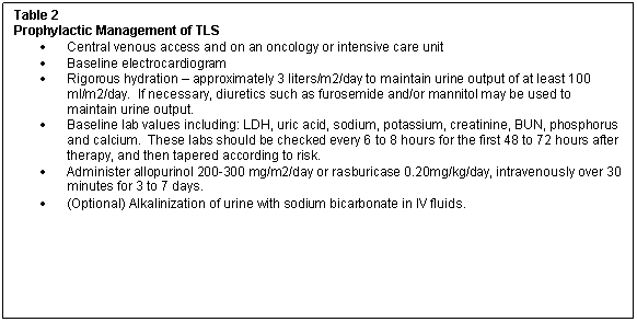
Alkalinization of urine, to a pH of 7-8, has long been a mainstay of preventive therapy because it promotes the excretion or uric acid in the urine. Recently, this has come under scrutiny, as over-alkalinization can actually promote calcium-phosphate crystallization. Many feel that by maintaining adequate urine output, calcium phosphate will be diluted, and the possibility of crystal formation will be low.
Allopurinol (Zyloprim®) works to block the development of new uric acid and to reduce the incidence of crystallization leading to obstruction, but cannot decrease uric acid formed before its initiation. It is known to commonly cause allergic reactions and is cleared by the kidney, so doses should be adjusted in renal failure. An intravenous form of allopurinol is also available (Aloprim™), which may be helpful in specific cases. Both are recommended to be started 24-48 hours before cytotxic therapy.
Rasburicase (Elitek™) is a relatively new intravenous agent used to lower both new and previously produced uric acid. Clinical trials found that uric acid levels decreased 85% with rasburicase, as compared with 12% with allopurinol, within 4 hours of starting the medications. This drug can cause anaphylactic reaction and nurses should have appropriate medications (diphenhydramine and epinephrine) at the bedside. Patients with G-6PD deficiency should not receive rasburicase because severe hemolysis can develop. Blood specimens for uric acid levels should be placed immediately on ice to prevent ex vivo breakdown of uric acid and inaccurate lab results.
Caution should be used in repleting electrolytes, as to not exacerbate pending TLS abnormalities.
Treatment
If electrolyte abnormalities develop, they can be managed as follows. Hyperkalemia should be treated quickly with sodium polystyrene sulfonate ( Kayexalate, Kionex ), 15-60 grams/day taken by mouth. Hyperphosphotemia is treated with aluminum hydroxide, 15 ml every 4 to 6 hours. In patients with asymptomatic hypocalcemia, treatment is not recommended due to an increase risk of calcium-phosphate precipitation. If the patient is symptomatic, calcium gluconate can be administered, but caution should be taken to monitor for worsening kidney function or decreased urine output.
For patients with severe abnormalities or acute renal failure, hemodialysis should be initiated emergently. This is a potentially reversible situation when treated promptly.
Nursing Interventions
Nursing interventions should include monitoring of urine output and alkalinization, when sodium bicarbonate is used. Any decrease in output should be reported; diuretics should be given and/or intravenous fluids should be adjusted accordingly. Patients must also be observed for fluid overload, which is accomplished by monitoring weight, output, vital signs and respiratory status. Nurses should monitor laboratory values and avoid using nephrotoxic medications (aminoglycoside antibiotics, NSAIDs). It may be necessary to institute a renal diet, which is low in potassium and phosphorus.
Education for the patient and family should include symptoms to be reported, a discussion of the patient's typical diet, and what foods should be avoided (those high in potassium & phosphorus; bananas, oranges, tomatoes, milk products, prepared/processed foods, sodas, chocolate and nuts). The patient should be encouraged to maintain adequate fluid intake and be aware of the need for accurate input and output monitoring. This can be a very scary time for patients, and thus the support of a knowledgeable nurse can make all the difference.
References
- Abeloff, M., Armitage, J., Niederhuber, J., Kastan, M. & McKenna, G. (Eds.): Clinical Oncology (2004). Elsevier, Philadelphia , PA.
- Cairo , M. S. and M. Bishop (2004). "Tumour lysis syndrome: new therapeutic strategies and classification." British Journal of Hematology 127 (1): 3-11.
- Cope, D. (2004). "Tumor lysis syndrome." Clincal Journal of Oncology Nursing 8 (4): 415-6.
- Doane, L. (2002). "Overview of tumor lysis syndrome." Seminars in Oncology Nursing 18 (3 Suppl 3): 2-5.
- Gobel, B. H. (2002). "Management of tumor lysis syndrome: prevention and treatment." Seminars in Oncology Nursing 18 (3 Suppl 3): 12-6.
- Krimsky, W. S., R. J. Behrens, et al. (2002). "Oncologic emergencies for the internist." Cleveland Clinic Journal of Medicine 69 (3): 209-10, 213-4, 216-7 passim.