The Pap Test: Cervical Changes and Further Testing
If you have had a Pap test that showed changes in the cells of your cervix, or a positive HPV test, this article will help you understand these test results and any other testing you may need.
What is a Pap Test and how is it done?
The Pap test, also called a "Pap smear," is used to find any changes in the cells of your cervix. During this test:
- A tool called a speculum is placed into the vagina so your provider can see your cervix and vagina.
- A cotton-tipped swab or small brush is used to take cells (also called a sample) from the cervix (area at the top of the vagina).
- The sample is sent to a lab. It is looked at under a microscope for abnormal cervical cells. These abnormal cells can become cancer.
- A report will be sent to your provider, who will talk with you about the results.
Normal discharge from your vagina has cells that are shed from the cervix and uterus. Samples of these cells are taken for the Pap test. For this reason, you should not douche, have vaginal intercourse, use tampons or vaginal medication for 48 hours before the Pap test is done.
The sample may be tested for Human Papilloma Virus (HPV) using a cotest. A cotest is both a HPV and Pap test. There are 14 types of HPV that are thought to be "high-risk" strains that can cause cervical cancer including strains 16 and 18. This test looks for these high-risk types of HPV in the cervical cells. This gives your provider more information about a possible cause of an abnormal pap result so you can plan the next steps.
When should cervical cancer screening be done?
The American Cancer Society has guidelines for cervical and HPV screening that are based on your age, screening history, risk factors, and the availability of screening tests (some centers do not have HPV only tests). If you have had your uterus and cervix removed in a hysterectomy and have no history of cervical cancer or pre-cancer, you should not be screened.
It is important to get screened, no matter which test you get. Even if you have had the HPV vaccine, you still need to be screened. If you are at high risk for cervical cancer, you may need to be screened more often. You might be high risk if you have an HIV infection, have had an organ transplant, or had in-utero exposure to the drug DES.
The American Cancer Society recommends the following guidelines for cervical cancer screening:
Ages 21-24
- No screening recommended.
Ages 25-29
- HPV test every 5 years (preferred); OR
- HPV/Pap cotest every 5 years (acceptable); OR
- Pap test every 3 years (acceptable).
Ages 30-65
- HPV test every 5 years (preferred); OR
- HPV/Pap cotest every 5 years (acceptable); OR
- Pap test every 3 years (acceptable).
Age 65 and older
- No screening if a series of prior tests were normal:
- Normal screening is having 2 negative HPV tests in a row, or 2 negative cotests/3 negative Pap tests in the past 10 years (with the most recent test in the past 3-5 years).
If you have had an abnormal or positive Pap or HPV test, you and your provider will talk about next steps. You may need more testing, a procedure, or treatment. Talk with your provider about what an abnormal test means for you and how you should be screened in the future.
What cervical changes can happen?
The cervix is made up of two parts:
- Ectocervix- The outer surface that opens into the vagina.
- Endocervix- The inner surface that lines the cervix (cervical canal).
These surfaces are covered by two types of cells:
- Squamous cells - Flat, thin cells that cover the ectocervix.
- Squamous cell carcinoma (SCC) - the most common cervical cancer type (about 70% of cases), starts in the squamous cells of the cervix.
- Glandular cells – Taller, column-shaped cells that cover the endocervix.
- Adenocarcinoma - a less common type (about 25% of cases) of cervical cancer, it starts in the glandular cells of the cervix. It can be harder to diagnose since it starts higher up in the cervix making these cells harder to find.
The area where the squamous cells and glandular cells meet is known as the “transformation zone.” This is where most cervical cancers start. The Pap test is taken from this area, since most dysplasia (see below) and cancers start here.
Two common changes in cells are metaplasia and dysplasia:
- Metaplasia - Metaplasia is cell growth or cell repair that is benign (not cancerous). This process normally occurs in unborn babies, during adolescence, and with the first pregnancy. Studies have shown that metaplasia is found in more than 50% of all women at some point. This is a normal finding and the changes are not cancer.
- Dysplasia - In dysplasia, there is an increase in the number of cells, but they do not mature as expected. This changes the inside of the cell. The higher the grade of dysplasia found on the cervix, the more likely that it will turn into invasive cancer. For this reason, dysplasia is thought of as a "pre-cancerous" condition. Dysplasias are almost always curable if treated. Although some mild dysplasias (such as LSIL) will get better without treatment, it is not possible to know which will get better on their own or may turn into cancer. For this reason, you may need more testing. Another name for cervical dysplasia is cervical intraepithelial neoplasia, or CIN.
What causes changes to cervical cells?
Inflammation often causes a mildly abnormal Pap test. An inflamed cervix may look red, irritated, or eroded. Some of the common causes of cervical inflammation are:
- Bacteria (from an infection), yeast or monilia infections, or trichomonas infections.
- Viruses, especially herpes infections and condyloma cuminata (warts).
- Pregnancy, miscarriage, or abortion.
- Chemicals (for example, medications).
- Hormonal changes.
When the inflammation is treated, natural repair of the tissues through metaplasia will often follow.
What do my Pap test results mean?
There are many possible results of your Pap test. The most common system for describing Pap test results is the Bethesda System (TBS). There are 3 main results:
- Negative for intraepithelial lesion or malignancy.
- Epithelial cell abnormalities.
- Other malignant neoplasms.
Some other information that may be included in your results:
Classification of Squamous Cells on the Pap Test (TBS)
- ASC - Atypical squamous cells. This is the most common abnormal finding in Pap tests. The Bethesda System divides ASC into two groups:
- ASC-US - Atypical squamous cells of undetermined significance. The squamous cells do not look completely normal, but it is uncertain what the cell changes mean. Sometimes the changes are related to HPV infection, but they can also be caused by other factors like pregnancy. If you are found to have ASC-US, your cells may then be tested for high-risk HPV.
- ASC-H - Atypical squamous cells, cannot exclude a high-grade squamous intraepithelial (cells on the surface of the cervix) lesion. ASC-H cells do not look normal, but it is uncertain what the cell changes mean.
- LSIL - Low-grade squamous intraepithelial lesion. This is the earliest pre-cancerous lesion. LSILs may be called mild dysplasia or cervical intraepithelial neoplasia type 1 (CIN-1).
- HSIL - High-grade squamous intraepithelial lesion. HSILs are more abnormal-looking than LSILs and have a higher chance of turning into cancer. May also be called moderate or severe dysplasia, carcinoma in situ, and/or CIN-2 and CIN-3.
- Squamous cell carcinoma is the most advanced category. This means that abnormal cervical squamous cells have invaded (spread) into the cervix. A finding of squamous cell carcinoma requires more testing and treatment. Keep in mind, when women undergo appropriate screening, most of the time, abnormalities in the cervix are detected and treated before they have had the chance to progress to cervical cancer.
Classification of Glandular Cells on the Pap Test
Glandular cells make mucus and are found in the opening of the cervix and in the uterus. Abnormalities in these cells are harder to classify. Glandular cells that are seen on the Pap test are often from the endocervix (area closest to the uterus). However, there are other areas that have glandular epithelial cells that may shed cells that can be seen on the Pap test. Endometrial cells may also be seen on Pap tests and can reveal other changes. Because the female reproductive tract is open to the abdominal cavity via the fallopian tubes, cells from the ovary, fallopian tubes, peritoneum, or other abdominal organs may be seen on the Pap smear. Glandular cells on the Pap test are classified as follows:
- Endometrial cells, cytologically benign, in a postmenopausal woman.
- Atypical glandular cells (AGC, formerly AGUS) that should be classified further, if possible, as to whether a reactive or neoplastic process is favored.
- Endocervical Adenocarcinoma.
- Endometrial Adenocarcinoma.
- Extrauterine Adenocarcinoma (e.g. ovarian, Fallopian tube, pancreas, etc.).
- Adenocarcinoma, not otherwise specified (i.e. unknown primary site).
What happens next after an abnormal Pap or HPV test?
An abnormal Pap or HPV test often calls for more testing, which often includes a biopsy. If the abnormality is minor (i.e. inflammation or HPV changes), your healthcare provider may choose to repeat the Pap test in a few months, as your own immune system may "clear" the HPV infection and a follow up Pap be normal. If the abnormalities do not clear or if they have gotten worse, you may need more testing. Your provider may send you first for a colposcopy:
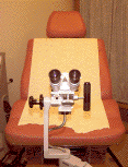
Colposcopy - A colposcope is a lighted microscope that is used to magnify the cervical tissue during a pelvic exam. The colposcope is used to see abnormal areas of the cervix and vagina that are too small to see with the naked eye. The whole transformation zone (described above) must be seen. The colposcopic exam is an office procedure and may be a bit more uncomfortable than a routine pelvic exam because of the pressure from the speculum lasting longer than a Pap test. The test takes 5 to 10 minutes to do.
During the exam, the provider may take small samples of cervical tissue (biopsies), which are later looked at by a pathologist under a microscope. These biopsies will guide further testing and/or treatment.
From the examiner's view
From the patient's view
Other biopsy and treatment options
Cone Biopsy: A cone biopsy (also called cold knife cone biopsy) is a minor operation, often done in an outpatient surgical facility. In the operating room, the physician removes a small cone-shaped tissue sample from your inner cervix. This tissue is sent to a pathologist to look at under a microscope. This procedure does not remove any of your reproductive organs and should have little impact on your future ability to become pregnant. If only dysplasia is found in the cone specimen, then often no additional treatment will be needed. However, if invasive cancer is found, more treatment (i.e. surgery or radiation therapy) may be needed. Therefore, a cone biopsy may be considered as therapeutic (if all of the dysplasia is removed) or diagnostic (if it discovers a worse problem that needs more treatment).
Loop Electrosurgical Excision Procedure (LEEP): The LEEP procedure is like a cone biopsy in that it removes a tissue sample from your cervix, which is then looked at under a microscope by a pathologist. It may also be called an LLETZ (large loop excision of the transformational zone). The LEEP procedure uses a low voltage, electric wire to cut away the abnormal area and can be done in the office with local anesthesia. However, the LEEP procedure and cone biopsy are not the same. Your provider will recommend which is the best option, depending on your case.
Cryosurgery - Cryosurgery is another treatment option that can be done in the doctor's office. During the procedure, the doctor freezes and destroys the dysplasia on your cervix. You may notice a brief unpleasant cold sensation during the freezing procedure. A disadvantage of cryosurgery is that no biopsy is taken to rule out the possibility of invasive cancer.
If you have questions about your Pap test or the results of your test, talk with your provider.
References
American Congress of Obstetricians and Gynecologists: Cervical Cancer Screening & Abnormal Cervical Cancer Screening Test Results.
Association of Reproductive Health Professionals - "Understanding Cervical Cancer Screening Test Results"
Centers for Disease Control "Cervical Cancer Screening"
National Institute of Health (2020). ACS’s Updated Cervical Cancer Screening Guidelines Explained. Taken from https://www.cancer.gov/news-events/cancer-currents-blog/2020/cervical-cancer-screening-hpv-test-guideline.
Saslow D, Solomon D, Lawson HW, et al. American Cancer Society, American Society for Colposcopy and Cervical Pathology, and American Society for Clinical Pathology screening guidelines for the prevention and early detection of cervical cancer. CA Cancer J Clin. 2012; 62: 147- 172.
World Health Organization. Human Papillomavirus and Cervical Cancer. 2019.
Wright T, Massad L, Dunton C, et al.2006 Consensus Guidelines for the Management of Women with Abnormal Cervical Cancer Screening Tests. American Journal of Obstetrics and Gynecology (2007;197(4):346-355).

