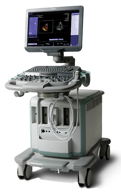
Ultrasound
An ultrasound (US) is a radiology test that makes pictures of organs inside your body. The ultrasound makes the pictures using sound waves. A probe is pressed against your skin and waves are created. As these sound waves bounce off your organ and return to the probe, an attached computer translates the pattern. The computer then uses the pattern to create an image that can be read by your provider. US waves work well in parts of the body made up of tissue or water. US does not work well in areas of the body where there is air or bone, such as the lungs or head.
Why are ultrasounds done?
Ultrasound is most often used to look at a growing baby. It can also be used to:
- See organs in the belly (liver, kidney, gallbladder).
- Look at the heart and blood vessels (arteries and veins).
- See lymph nodes.
- Find and see a mass more clearly during a biopsy.
- See how much urine is in your bladder.
Ultrasounds can be used in emergency situations, looking for blood in the belly, a blood clot in the leg, or any changes or problems in the heart. Most often ultrasounds are done non-emergently and scheduled as an outpatient procedure. They can be used with a physical exam and lab work to diagnose a medical problem.
How do I prepare for an ultrasound?
How you prepare for an ultrasound depends on the part of your body that needs testing. In some cases, you don’t need to prepare. Other times, you may be asked not to eat or drink for up to 12 hours before the test, or you may need to drink up to six glasses of water two hours before the test.
If the US will be placed rectally for a prostate biopsy, you may need to clear stool from your bowel. If the scope will be inserted either endoscopically (through the mouth and esophagus) or bronchoscopically (into the lungs), you will be asked to fast (not eat) to prepare for the sedation medications.
Your provider will give you instructions on how to prepare for your US.
How is this test done?
You will likely be asked to put on a hospital gown. The position you will lie in on the table depends on the part of your body being looked at. Gel will be placed on your skin to allow the probe to make better contact with your skin. The technician will then place a transducer or probe on your skin above the area being looked at. The probe makes sound waves that travel into the tissue and then reflect off tissues inside your body and return to the probe.
The test may cause discomfort in some cases but not pain. It only takes 20-40 minutes to complete, depending on the size of the area being looked at.
How do I get the results of my ultrasound?
The radiologist (a doctor who specializes in medical imaging) writes a report for your healthcare provider who ordered the ultrasound. The report will give both normal and abnormal findings. Your provider will be able to discuss your results with you.
References
U.S. Department of Health and Human Services. (2016, July). Ultrasound. National Institute of Biomedical Imaging and Bioengineering. Retrieved September 15, 2022, from https://www.nibib.nih.gov/science-education/science-topics/ultrasound
