National Cancer Institute
Post Date: Oct 10, 2023
Non-small cell lung cancer (NSCLC) treatment options include surgery, chemotherapy, radiation therapy, targeted therapy, and immunotherapy. Laser therapy, photodynamic therapy, cryosurgery, and electrocautery may be used. Learn more about NSCLC in this expert-reviewed summary.
Non-Small Cell Lung Cancer Treatment
General Information About Non-Small Cell Lung Cancer
Key Points for this Section
- Non-small cell lung cancer is a disease in which malignant (cancer) cells form in the tissues of the lung.
- There are several types of non-small cell lung cancer.
- Smoking is the major risk factor for non-small cell lung cancer.
- Signs of non-small cell lung cancer include a cough that doesn't go away and shortness of breath.
- Tests that examine the lungs are used to diagnose and stage non-small cell lung cancer.
- If lung cancer is suspected, a biopsy is done.
- Certain factors affect prognosis (chance of recovery) and treatment options.
- For most patients with non-small cell lung cancer, current treatments do not cure the cancer.
Non-small cell lung cancer is a disease in which malignant (cancer) cells form in the tissues of the lung.
The lungs are a pair of cone-shaped organs in the chest. The lungs bring oxygen into the body as you breathe in. They release carbon dioxide, a waste product of the body's cells, as you breathe out. Each lung has sections called lobes. The left lung has two lobes. The right lung is slightly larger and has three lobes. Two tubes called bronchi lead from the trachea (windpipe) to the right and left lungs. The bronchi are sometimes also involved in lung cancer. Tiny air sacs called alveoli and small tubes called bronchioles make up the inside of the lungs.
 Anatomy of the respiratory system showing the trachea, the right and left lungs and their lobes, and the bronchi. The lymph nodes and the diaphragm are also shown. Oxygen is inhaled into the lungs and passes through the alveoli (the tiny air sacs at the end of the bronchioles) and into the bloodstream (see inset), where it travels to the tissues throughout the body.
A thin membrane called the pleura covers the outside of each lung and lines the inside wall of the chest cavity. This creates a sac called the pleural cavity. The pleural cavity normally contains a small amount of fluid that helps the lungs move smoothly in the chest when you breathe.
There are two main types of lung cancer: non-small cell lung cancer and small cell lung cancer.
There are several types of non-small cell lung cancer.
Each type of non-small cell lung cancer has different kinds of cancer cells. The cancer cells of each type grow and spread in different ways. The types of non-small cell lung cancer are named for the kinds of cells found in the cancer and how the cells look under a microscope:
- Squamous cell carcinoma: Cancer that forms in the thin, flat cells lining the inside of the lungs. This is also called epidermoid carcinoma.
- Large cell carcinoma: Cancer that may begin in several types of large cells.
- Adenocarcinoma: Cancer that begins in the cells that line the alveoli and make substances such as mucus.
Other less common types of non-small cell lung cancer are: adenosquamous carcinoma, sarcomatoid carcinoma, salivary gland carcinoma, carcinoid tumor, and unclassified carcinoma.
Smoking is the major risk factor for non-small cell lung cancer.
lung cancerAnything that increases your chance of getting a disease is called a risk factor. Having a risk factor does not mean that you will get cancer; not having risk factors doesn't mean that you will not get cancer. Talk to your doctor if you think you may be at risk for lung cancer.
Risk factors for lung cancer include the following:
- Smoking cigarettes, pipes, or cigars, now or in the past. This is the most important risk factor for lung cancer. The earlier in life a person starts smoking, the more often a person smokes, and the more years a person smokes, the greater the risk of lung cancer.
- Being exposed to secondhand smoke.
- Being exposed to asbestos, arsenic, chromium, beryllium, nickel, soot, or tar in the workplace.
- Being exposed to radiation from any of the following:
- Radiation therapy to the breast or chest.
- Radon in the home or workplace.
- Imaging tests such as CT scans.
- Atomic bomb radiation.
- Living where there is air pollution.
- Having a family history of lung cancer.
- Being infected with the human immunodeficiency virus (HIV).
- Taking beta carotenesupplements and being a heavy smoker.
Older age is the main risk factor for most cancers. The chance of getting cancer increases as you get older.
When smoking is combined with other risk factors, the risk of lung cancer is increased.
Signs of non-small cell lung cancer include a cough that doesn't go away and shortness of breath.
Sometimes lung cancer does not cause any signs or symptoms. It may be found during a chest x-ray done for another condition. Signs and symptoms may be caused by lung cancer or by other conditions. Check with your doctor if you have any of the following:
- Chest discomfort or pain.
- A cough that doesn’t go away or gets worse over time.
- Trouble breathing.
- Wheezing.
- Blood in sputum (mucus coughed up from the lungs).
- Hoarseness.
- Loss of appetite.
- Weight loss for no known reason.
- Feeling very tired.
- Trouble swallowing.
- Swelling in the face and/or veins in the neck.
Tests that examine the lungs are used to diagnose and stage non-small cell lung cancer.
Tests and procedures to diagnose and stage non-small cell lung cancer are often done at the same time. In addition to asking about your personal and family health history and doing a physical exam, your doctor may perform the following tests and procedures:
- Laboratory tests: Medical procedures that test samples of tissue, blood, urine, or other substances in the body. These tests help to diagnose disease, plan and check treatment, or monitor the disease over time.
- Chest x-ray: An x-ray of the organs and bones inside the chest. An x-ray is a type of energy beam that can go through the body and onto film, making a picture of areas inside the body.
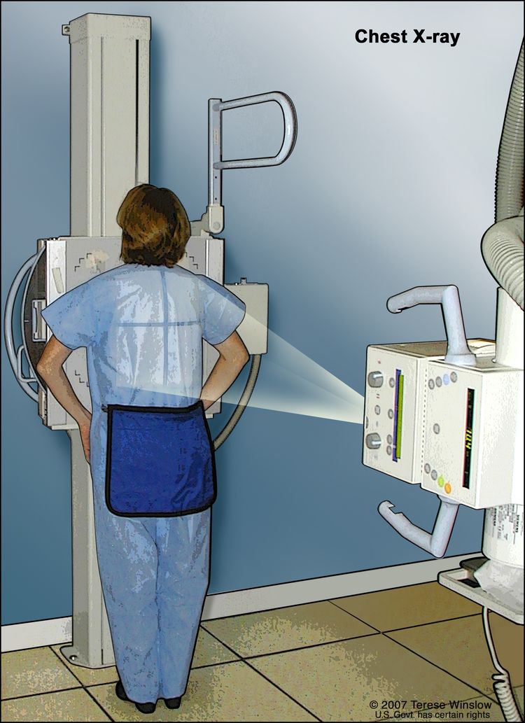 A chest x-ray is used to take pictures of the structures and organs inside the chest. X-rays pass through the patient's body onto film or a computer.
 - CT scan (CAT scan): A procedure that makes a series of detailed pictures of areas inside the body, such as the chest, taken from different angles. The pictures are made by a computer linked to an x-ray machine. A dye may be injected into a vein or swallowed to help the organs or tissues show up more clearly. This procedure is also called computed tomography, computerized tomography, or computerized axial tomography.
- Sputum cytology: A procedure in which a pathologist views a sample of sputum (mucus coughed up from the lungs) under a microscope, to check for cancer cells.
- Thoracentesis: The removal of fluid from the space between the lining of the chest and the lung, using a needle. A pathologist views the fluid under a microscope to look for cancer cells.
If lung cancer is suspected, a biopsy is done.
One of the following types of biopsies is usually used:
- Fine-needle aspiration (FNA) biopsy of the lung: The removal of tissue or fluid from the lung using a thin needle. A CT scan, ultrasound, or other imaging procedure is used to locate the abnormal tissue or fluid in the lung. A small incision may be made in the skin where the biopsy needle is inserted into the abnormal tissue or fluid. A sample is removed with the needle and sent to the laboratory. A pathologist then views the sample under a microscope to look for cancer cells. A chest x-ray is done after the procedure to make sure no air is leaking from the lung into the chest.
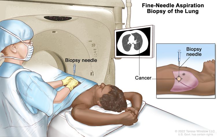 Fine-needle aspiration biopsy of the lung. The patient lies on a table that slides through the computed tomography (CT) machine, which takes x-ray pictures of the inside of the body. The x-ray pictures help the doctor see where the abnormal tissue is in the lung. A biopsy needle is inserted through the chest wall and into the area of abnormal lung tissue. A small piece of tissue is removed through the needle and checked under the microscope for signs of cancer.
 An endoscopic ultrasound (EUS) is a type of ultrasound that may be used to guide an FNA biopsy of the lung, lymph nodes, or other areas. EUS is a procedure in which an endoscope is inserted into the body. An endoscope is a thin, tube-like instrument with a light and a lens for viewing. A probe at the end of the endoscope is used to bounce high-energy sound waves (ultrasound) off internal tissues or organs and make echoes. The echoes form a picture of body tissues called a sonogram.
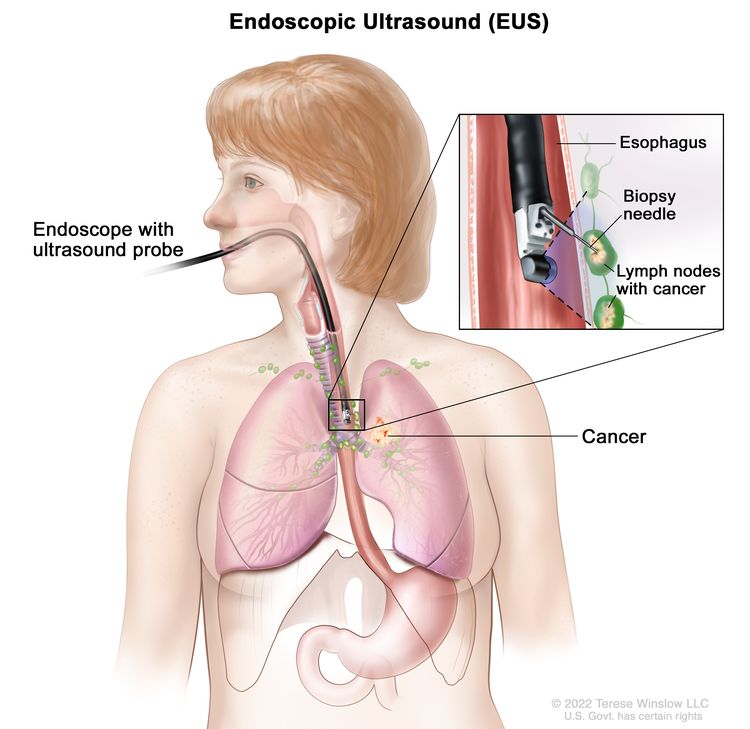 Endoscopic ultrasound-guided fine-needle aspiration biopsy. An endoscope that has an ultrasound probe and a biopsy needle is inserted through the mouth and into the esophagus. The probe bounces sound waves off body tissues to make echoes that form a sonogram (computer picture) of the lymph nodes near the esophagus. The sonogram helps the doctor see where to place the biopsy needle to remove tissue from the lymph nodes. This tissue is checked under a microscope for signs of cancer.
 - Bronchoscopy: A procedure to look inside the trachea and large airways in the lung for abnormal areas. A bronchoscope is inserted through the nose or mouth into the trachea and lungs. A bronchoscope is a thin, tube-like instrument with a light and a lens for viewing. It may also have a tool to remove tissue samples, which are checked under a microscope for signs of cancer.
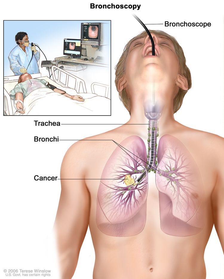 Bronchoscopy. A bronchoscope is inserted through the mouth, trachea, and major bronchi into the lung, to look for abnormal areas. A bronchoscope is a thin, tube-like instrument with a light and a lens for viewing. It may also have a cutting tool. Tissue samples may be taken to be checked under a microscope for signs of disease.
 - Thoracoscopy: A surgical procedure to look at the organs inside the chest to check for abnormal areas. An incision (cut) is made between two ribs, and a thoracoscope is inserted into the chest. A thoracoscope is a thin, tube-like instrument with a light and a lens for viewing. It may also have a tool to remove tissue or lymph node samples, which are checked under a microscope for signs of cancer. In some cases, this procedure is used to remove part of the esophagus or lung. If certain tissues, organs, or lymph nodes can’t be reached, a thoracotomy may be done. In this procedure, a larger incision is made between the ribs and the chest is opened.
- Mediastinoscopy: A surgical procedure to look at the organs, tissues, and lymph nodes between the lungs for abnormal areas. An incision (cut) is made at the top of the breastbone and a mediastinoscope is inserted into the chest. A mediastinoscope is a thin, tube-like instrument with a light and a lens for viewing. It may also have a tool to remove tissue or lymph node samples, which are checked under a microscope for signs of cancer.
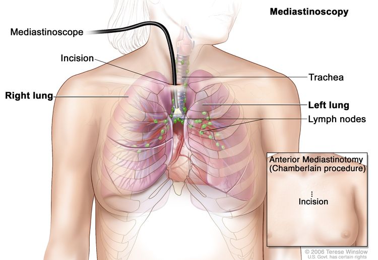 Mediastinoscopy. A mediastinoscope is inserted into the chest through an
incision above the breastbone to look for abnormal areas between the
lungs. A mediastinoscope is a thin, tube-like instrument with a light
and a lens for viewing. It may also have a cutting tool. Tissue samples
may be taken from lymph nodes on the right side of the chest and checked
under a microscope for signs of cancer. In an anterior mediastinotomy
(Chamberlain procedure), the incision is made beside the breastbone to
remove tissue samples from the lymph nodes on the left side of the
chest.
Mediastinoscopy. A mediastinoscope is inserted into the chest through an
incision above the breastbone to look for abnormal areas between the
lungs. A mediastinoscope is a thin, tube-like instrument with a light
and a lens for viewing. It may also have a cutting tool. Tissue samples
may be taken from lymph nodes on the right side of the chest and checked
under a microscope for signs of cancer. In an anterior mediastinotomy
(Chamberlain procedure), the incision is made beside the breastbone to
remove tissue samples from the lymph nodes on the left side of the
chest.
- Anterior mediastinotomy: A surgical procedure to look at the organs and tissues between the lungs and between the breastbone and heart for abnormal areas. An incision (cut) is made next to the breastbone and a mediastinoscope is inserted into the chest. A mediastinoscope is a thin, tube-like instrument with a light and a lens for viewing. It may also have a tool to remove tissue or lymph node samples, which are checked under a microscope for signs of cancer. This is also called the Chamberlain procedure.
- Lymph node biopsy: The removal of all or part of a lymph node. A pathologist views the lymph node tissue under a microscope to check for cancer cells.
One or more of the following laboratory tests may be done to study the tissue samples:
- Molecular test: A laboratory test to check for certain genes, proteins, or other molecules in a sample of tissue, blood, or other body fluid. Molecular tests check for certain gene or chromosome changes that occur in non-small cell lung cancer.
- Immunohistochemistry: A laboratory test that uses antibodies to check for certain antigens (markers) in a sample of a patient’s tissue. The antibodies are usually linked to an enzyme or a fluorescent dye. After the antibodies bind to a specific antigen in the tissue sample, the enzyme or dye is activated, and the antigen can then be seen under a microscope. This type of test is used to help diagnose cancer and to help tell one type of cancer from another type of cancer.
Certain factors affect prognosis (chance of recovery) and treatment options.
The prognosis and treatment options depend on the following:
- The stage of the cancer (the size of the tumor and whether it is in the lung only or has spread to other places in the body).
- The type of lung cancer.
- Whether the cancer has mutations (changes) in certain genes, such as the epidermal growth factor receptor (EGFR) gene or the anaplastic lymphoma kinase (ALK) gene.
- Whether there are signs and symptoms such as coughing or trouble breathing.
- The patient’s general health.
For most patients with non-small cell lung cancer, current treatments do not cure the cancer.
If lung cancer is found, taking part in one of the many clinical trials being done to improve treatment should be considered. Clinical trials are taking place in most parts of the country for patients with all stages of non-small cell lung cancer. Information about ongoing clinical trials is available from the NCI website.
Stages of Non-Small Cell Lung Cancer
Key Points for this Section
- After lung cancer has been diagnosed, tests are done to find out if cancer cells have spread within the lungs or to other parts of the body.
- There are three ways that cancer spreads in the body.
- Cancer may spread from where it began to other parts of the body.
- The following stages are used for non-small cell lung cancer:
- Occult (hidden) stage
- Stage 0
- Stage I
- Stage II
- Stage III
- Stage IV
- Non-small cell lung cancer can recur (come back) after it has been treated.
After lung cancer has been diagnosed, tests are done to find out if cancer cells have spread within the lungs or to other parts of the body.
The process used to find out if cancer has spread within the lungs or to other parts of the body is called staging. The information gathered from the staging process determines the stage of the disease. It is important to know the stage in order to plan treatment. Some of the tests used to diagnose non-small cell lung cancer are also used to stage the disease. For more information, see the General Information section.
Other tests and procedures that may be used in the staging process include the following:
- MRI (magnetic resonance imaging): A procedure that uses a magnet, radio waves, and a computer to make a series of detailed pictures of areas inside the body, such as the brain. This procedure is also called nuclear magnetic resonance imaging (NMRI).
- CT scan (CAT scan): A procedure that makes a series of detailed pictures of areas inside the body, such as the brain, abdomen, and lymph nodes, taken from different angles. The pictures are made by a computer linked to an x-ray machine. A dye may be injected into a vein or swallowed to help the organs or tissues show up more clearly. This procedure is also called computed tomography, computerized tomography, or computerized axial tomography.
- PET scan (positron emission tomography scan): A procedure to find cancer cells in the body. A small amount of radioactiveglucose (sugar) is injected into a vein. The PET scanner rotates around the body and makes a picture of where glucose is being used in the body. Malignant tumor cells show up brighter in the picture because they are more active and take up more glucose than normal cells do.
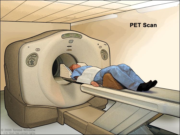 PET (positron emission tomography) scan. The patient lies on a table that slides through the PET machine. The head rest and white strap help the patient lie still. A small amount of radioactive glucose (sugar) is injected into the patient's vein, and a scanner makes a picture of where the glucose is being used in the body. Cancer cells show up brighter in the picture because they take up more glucose than normal cells do.
- Bone scan: A procedure to check if there are rapidly dividing cells, such as cancer cells, in the bone. A very small amount of radioactive material is injected into a vein and travels through the bloodstream. The radioactive material collects in the bones with cancer and is detected by a scanner.
- Pulmonary function test (PFT): A test to see how well the lungs are working. It measures how much air the lungs can hold and how quickly air moves into and out of the lungs. It also measures how much oxygen is used and how much carbon dioxide is given off during breathing. This is also called lung function test.
- Bone marrow aspiration and biopsy: The removal of bone marrow, blood, and a small piece of bone by inserting a hollow needle into the hipbone or breastbone. A pathologist views the bone marrow, blood, and bone under a microscope to look for signs of cancer.
There are three ways that cancer spreads in the body.
Cancer can spread through tissue, the lymph system, and the blood:
- Tissue. The cancer spreads from where it began by growing into nearby areas.
- Lymph system. The cancer spreads from where it began by getting into the lymph system. The cancer travels through the lymph vessels to other parts of the body.
- Blood. The cancer spreads from where it began by getting into the blood. The cancer travels through the blood vessels to other parts of the body.
Cancer may spread from where it began to other parts of the body.
When cancer spreads to another part of the body, it is called metastasis. Cancer cells break away from where they began (the primary tumor) and travel through the lymph system or blood.
- Lymph system. The cancer gets into the lymph system, travels through the lymph vessels, and forms a tumor (metastatic tumor) in another part of the body.
- Blood. The cancer gets into the blood, travels through the blood vessels, and forms a tumor (metastatic tumor) in another part of the body.
The metastatic tumor is the same type of cancer as the primary tumor. For example, if non-small cell lung cancer spreads to the brain, the cancer cells in the brain are actually lung cancer cells. The disease is metastatic lung cancer, not brain cancer.
metastasis: how cancer spreadsMany cancer deaths are caused when cancer moves from the original tumor and spreads to other tissues and organs. This is called metastatic cancer. This animation shows how cancer cells travel from the place in the body where they first formed to other parts of the body.The following stages are used for non-small cell lung cancer:
Occult (hidden) stage
In the occult (hidden) stage, cancer cannot be seen by imaging or bronchoscopy. Cancer cells are found in sputum or bronchial washings (a sample of cells taken from inside the airways that lead to the lungs). Cancer may have spread to other parts of the body.
Stage 0
In stage 0, abnormal cells are found in the lining of the airways. These abnormal cells may become cancer and spread into nearby normal tissue. Stage 0 may be adenocarcinoma in situ (AIS) or squamous cell carcinoma in situ (SCIS).
Stage I
In stage I, cancer has formed. Stage I is divided into stages IA and IB.
- Stage IA:
 Stage IA lung cancer. The tumor is in the lung only and is 3 centimeters or smaller. Cancer has not spread to the lymph nodes.
The tumor is in the lung only and is 3 centimeters or smaller. Cancer has not spread to the lymph nodes.
- Stage IB:
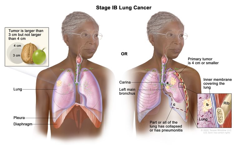 Stage IB lung cancer. The tumor is larger than 3 centimeters but not larger than 4 centimeters. Cancer has not spread to the lymph nodes; OR the tumor is 4 centimeters or smaller. Cancer has not spread to the lymph nodes and one or more of the following is found: (a) cancer has spread to the main bronchus, but has not spread to the carina; and/or (b) cancer has spread to the inner membrane that covers the lung; and/or (c) part of the lung or the whole lung has collapsed or has pneumonitis (inflammation of the lung).
The tumor is larger than 3 centimeters but not larger than 4 centimeters. Cancer has not spread to the lymph nodes.
or
The tumor is 4 centimeters or smaller and one or more of the following is found:
- Cancer has spread to the main bronchus, but has not spread to the carina.
- Cancer has spread to the innermost layer of the membrane that covers the lung.
- Part of the lung or the whole lung has collapsed or has developed pneumonitis.
Cancer has not spread to the lymph nodes.
Stage II
Stage II is divided into stages IIA and IIB.
- Stage IIA:
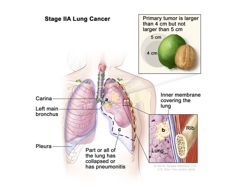 Stage IIA lung cancer. The tumor is larger than 4 centimeters but not larger than 5 centimeters. Cancer has not spread to the lymph nodes and one or more of the following may be found: (a) cancer has spread to the main bronchus, but has not spread to the carina; and/or (b) cancer has spread to the inner membrane that covers the lung; and/or (c) part of the lung or the whole lung has collapsed or has pneumonitis (inflammation of the lung).
The tumor is larger than 4 centimeters but not larger than 5 centimeters. Cancer has not spread to the lymph nodes and one or more of the following may be found:
- Cancer has spread to the main bronchus, but has not spread to the carina.
- Cancer has spread to the innermost layer of the membrane that covers the lung.
- Part of the lung or the whole lung has collapsed or has developed pneumonitis.
- Stage IIB:
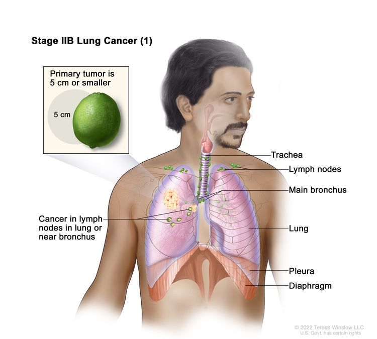 Stage IIB lung cancer (1). The primary tumor is 5 centimeters or smaller and cancer has spread to the lymph nodes on the same side of the chest as the primary tumor. The lymph nodes with cancer are in the lung or near the bronchus.
The tumor is 5 centimeters or smaller and cancer has spread to lymph nodes on the same side of the chest as the primary tumor. The lymph nodes with cancer are in the lung or near the bronchus. Also, one or more of the following may be found:
- Cancer has spread to the main bronchus, but has not spread to the carina.
- Cancer has spread to the innermost layer of the membrane that covers the lung.
- Part of the lung or the whole lung has collapsed or has developed pneumonitis.
or
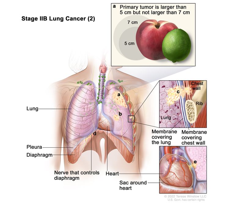 Stage IIB lung cancer (2). Cancer has not spread to lymph nodes and one or more of the following is found: (a) the primary tumor is larger than 5 centimeters but not larger than 7 centimeters; and/or (b) there are one or more separate tumors in the same lobe of the lung as the primary tumor; and/or cancer has spread to any of the following: (c) the chest wall and/or the membrane that lines the inside of the chest wall, (d) the nerve that controls the diaphragm, and/or (e) the outer layer of tissue of the sac around the heart.
Cancer has not spread to the lymph nodes and one or more of the following is found:
- The tumor is larger than 5 centimeters but not larger than 7 centimeters.
- There are one or more separate tumors in the same lobe of the lung as the primary tumor.
- Cancer has spread to any of the following:
- The membrane that lines the inside of the chest wall.
- Chest wall.
- The nerve that controls the diaphragm.
- Outer layer of tissue of the sac around the heart.
Stage III
Stage III is divided into stages IIIA, IIIB, and IIIC.
- Stage IIIA:
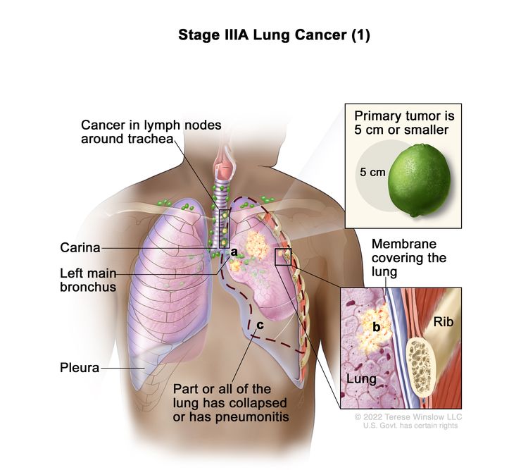 Stage IIIA lung cancer (1). The tumor is 5 centimeters or smaller and cancer has spread to lymph nodes on the same side of the chest as the primary tumor. The lymph nodes with cancer are around the trachea or aorta (not shown), or where the trachea divides into the bronchi. Also, one or more of the following may be found: (a) cancer has spread to the main bronchus, but has not spread to the carina; and/or (b) cancer has spread to the inner membrane that covers the lung; and/or (c) part of the lung or the whole lung has collapsed or has pneumonitis (inflammation of the lung).
The tumor is 5 centimeters or smaller and cancer has spread to lymph nodes on the same side of the chest as the primary tumor. The lymph nodes with cancer are around the trachea or aorta, or where the trachea divides into the bronchi. Also, one or more of the following may be found:
- Cancer has spread to the main bronchus, but has not spread to the carina.
- Cancer has spread to the innermost layer of the membrane that covers the lung.
- Part of the lung or the whole lung has collapsed or has developed pneumonitis.
or
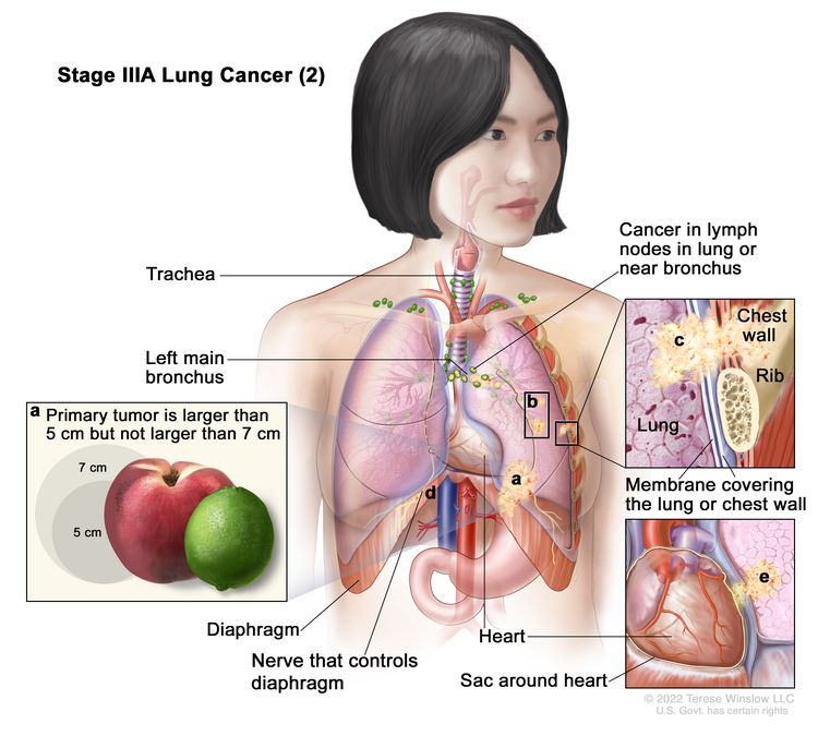 Stage IIIA lung cancer (2). Cancer has spread to lymph nodes on the same side of the chest as the primary tumor. The lymph nodes with cancer are in the lung or near the bronchus. Also, one or more of the following is found: (a) the tumor is larger than 5 centimeters but not larger than 7 centimeters; and/or (b) there are one or more separate tumors in the same lobe of the lung as the primary tumor; and/or cancer has spread to any of the following: (c) the chest wall and/or the membrane that lines the inside of the chest wall, (d) the nerve that controls the diaphragm, and/or (e) the outer layer of tissue of the sac around the heart.
Cancer has spread to lymph nodes on the same side of the chest as the primary tumor. The lymph nodes with cancer are in the lung or near the bronchus. Also, one or more of the following is found:
- The tumor is larger than 5 centimeters but not larger than 7 centimeters.
- There are one or more separate tumors in the same lobe of the lung as the primary tumor.
- Cancer has spread to any of the following:
- The membrane that lines the inside of the chest wall.
- Chest wall.
- The nerve that controls the diaphragm.
- Outer layer of tissue of the sac around the heart.
or
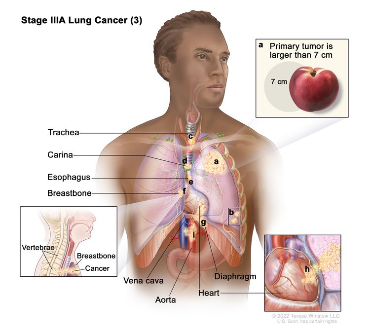 Stage IIIA lung cancer (3). Cancer may have spread to lymph nodes on the same side of the chest as the primary tumor. The lymph nodes with cancer are in the lung or near the bronchus. Also, one or more of the following is found: (a) the primary tumor is larger than 7 centimeters; and/or (b) there are one or more separate tumors in a different lobe of the lung with the primary tumor; and/or the tumor is any size and cancer has spread to any of the following: (c) trachea, (d) carina, (e) esophagus, (f) breastbone or backbone, (g) diaphragm, (h) heart, (i) major blood vessels that lead to or from the heart (aorta or vena cava), or the nerve that controls the larynx (not shown).
Cancer may have spread to lymph nodes on the same side of the chest as the primary tumor. The lymph nodes with cancer are in the lung or near the bronchus. Also, one or more of the following is found:
- The tumor is larger than 7 centimeters.
- There are one or more separate tumors in a different lobe of the lung with the primary tumor.
- The tumor is any size and cancer has spread to any of the following:
- Trachea.
- Carina.
- Esophagus.
- Breastbone or backbone.
- Diaphragm.
- Heart.
- Major blood vessels that lead to or from the heart (aorta or vena cava).
- Nerve that controls the larynx (voice box).
- Stage IIIB:
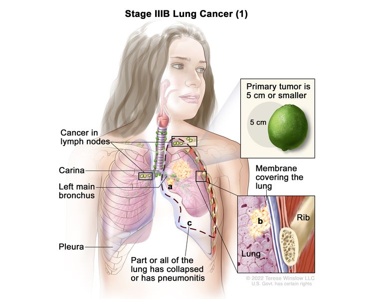 Stage IIIB lung cancer (1). The primary tumor is 5 centimeters or smaller and cancer has spread to lymph nodes above the collarbone on the same side of the chest as the primary tumor or to any lymph nodes on the opposite side of the chest as the primary tumor. Also, one or more of the following may be found: (a) cancer has spread to the main bronchus, but has not spread to the carina; and/or (b) cancer has spread to the inner membrane that covers the lung; and/or (c) part of the lung or the whole lung has collapsed or has pneumonitis (inflammation of the lung).
The tumor is 5 centimeters or smaller and cancer has spread to lymph nodes above the collarbone on the same side of the chest as the primary tumor or to any lymph nodes on the opposite side of the chest as the primary tumor. Also, one or more of the following may be found:
- Cancer has spread to the main bronchus, but has not spread to the carina.
- Cancer has spread to the innermost layer of the membrane that covers the lung.
- Part of the lung or the whole lung has collapsed or has developed pneumonitis.
or
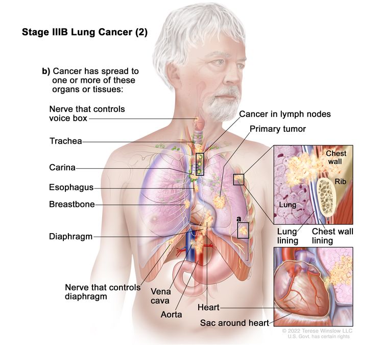 Stage IIIB lung cancer (2). The tumor may be any size and cancer has spread to lymph nodes on the same side of the chest as the primary tumor. The lymph nodes with cancer are around the trachea or aorta (not shown), or where the trachea divides into the bronchi. Also, one or more of the following is found: (a) there are one or more separate tumors in the same lobe or a different lobe of the lung with the primary tumor; and/or (b) cancer has spread to any of the following: the chest wall or the membrane that lines the inside of the chest wall, the nerve that controls the voice box, the trachea, the carina, the esophagus, the breastbone or backbone (not shown), the diaphragm, the nerve that controls the diaphragm, the heart, the major blood vessels that lead to or from the heart (aorta or vena cava), or the outer layer of tissue of the sac around the heart.
The tumor may be any size and cancer has spread to lymph nodes on the same side of the chest as the primary tumor. The lymph nodes with cancer are around the trachea or aorta, or where the trachea divides into the bronchi. Also, one or more of the following is found:
- There are one or more separate tumors in the same lobe or a different lobe of the lung with the primary tumor.
- Cancer has spread to any of the following:
- The membrane that lines the inside of the chest wall.
- Chest wall.
- The nerve that controls the diaphragm.
- Outer layer of tissue of the sac around the heart.
- Trachea.
- Carina.
- Esophagus.
- Breastbone or backbone.
- Diaphragm.
- Heart.
- Major blood vessels that lead to or from the heart (aorta or vena cava).
- Nerve that controls the larynx (voice box).
- Stage IIIC:
 Stage IIIC lung cancer. The tumor may be any size and cancer has spread to lymph nodes above the collarbone on the same side of the chest as the primary tumor or to any lymph nodes on the opposite side of the chest as the primary tumor. Also, one or more of the following is found: (a) there are one or more separate tumors in the same lobe or a different lobe of the lung with the primary tumor; and/or (b) cancer has spread to any of the following: the chest wall or the membrane that lines the inside of the chest wall, the nerve that controls the voice box, the trachea, the carina, the esophagus, the breastbone or backbone (not shown), the diaphragm, the nerve that controls the diaphragm, the heart, the major blood vessels that lead to or from the heart (aorta or vena cava), or the outer layer of tissue of the sac around the heart.
The tumor may be any size and cancer has spread to lymph nodes above the collarbone on the same side of the chest as the primary tumor or to any lymph nodes on the opposite side of the chest as the primary tumor. Also, one or more of the following is found:
- There are one or more separate tumors in the same lobe or a different lobe of the lung with the primary tumor.
- Cancer has spread to any of the following:
- The membrane that lines the inside of the chest wall.
- Chest wall.
- The nerve that controls the diaphragm.
- Outer layer of tissue of the sac around the heart.
- Trachea.
- Carina.
- Esophagus.
- Breastbone or backbone.
- Diaphragm.
- Heart.
- Major blood vessels that lead to or from the heart (aorta or vena cava).
- Nerve that controls the larynx (voice box).
Stage IV
Stage IV is divided into stages IVA and IVB.
- Stage IVA:
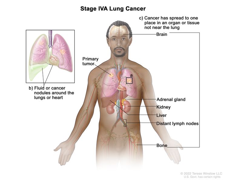 Stage IVA lung cancer. The tumor may be any size and cancer may have spread to the lymph nodes. One or more of the following is found: (a) there are one or more tumors in the lung that does not have the primary tumor; and/or (b) cancer is found in fluid around the lungs or heart or there are cancer nodules in the lining around the lungs or the sac around the heart; and/or (c) cancer has spread to one place in an organ or tissue not near the lung, such as the brain, adrenal gland, kidney, liver, or bone, or to a lymph node that is not near the lung.
The tumor may be any size and cancer may have spread to the lymph nodes. One or more of the following is found:
- There are one or more tumors in the lung that does not have the primary tumor.
- Cancer is found in the lining around the lungs or the sac around the heart.
- Cancer is found in fluid around the lungs or the heart.
- Cancer has spread to one place in an organ not near the lung, such as the brain, liver, adrenal gland, kidney, bone, or to a lymph node that is not near the lung.
- Stage IVB:
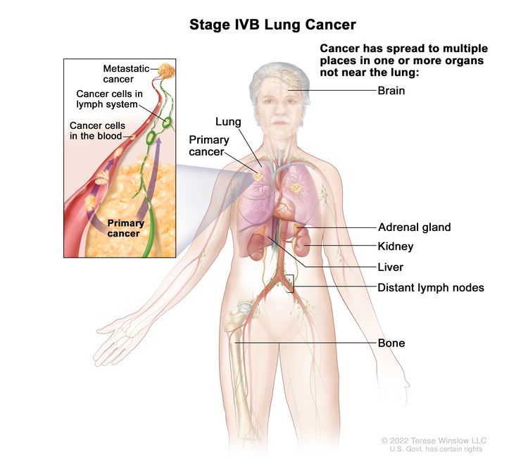 Stage IVB lung cancer. The cancer has spread to multiple places in one or more organs that are not near the lung, such as the brain, adrenal gland, kidney, liver, distant lymph nodes, or bone.
Cancer has spread to multiple places in one or more organs that are not near the lung.
Non-small cell lung cancer can recur (come back) after it has been treated.
The cancer may come back in the brain, lung, or other parts of the body.
Treatment Option Overview
Key Points for this Section
- There are different types of treatment for patients with non-small cell lung cancer.
- The following types of treatment are used:
- Surgery
- Radiation therapy
- Chemotherapy
- Targeted therapy
- Immunotherapy
- Laser therapy
- Photodynamic therapy (PDT)
- Cryosurgery
- Electrocautery
- New types of treatment are being tested in clinical trials.
- Radiosensitizers
- New combinations
- Treatment for non-small cell lung cancer may cause side effects.
- Patients may want to think about taking part in a clinical trial.
- Patients can enter clinical trials before, during, or after starting their cancer treatment.
- Follow-up tests may be needed.
There are different types of treatment for patients with non-small cell lung cancer.
Different types of treatments are available for patients with non-small cell lung cancer. Some treatments are standard (the currently used treatment), and some are being tested in clinical trials. A treatment clinical trial is a research study meant to help improve current treatments or obtain information on new treatments for patients with cancer. When clinical trials show that a new treatment is better than the standard treatment, the new treatment may become the standard treatment. Patients may want to think about taking part in a clinical trial. Some clinical trials are open only to patients who have not started treatment.
The following types of treatment are used:
Surgery
Four types of surgery are used to treat lung cancer:
- Wedge resection:
Surgery to remove a tumor and some of the normal tissue around it. When a slightly larger amount of tissue is taken, it is called a segmental resection.
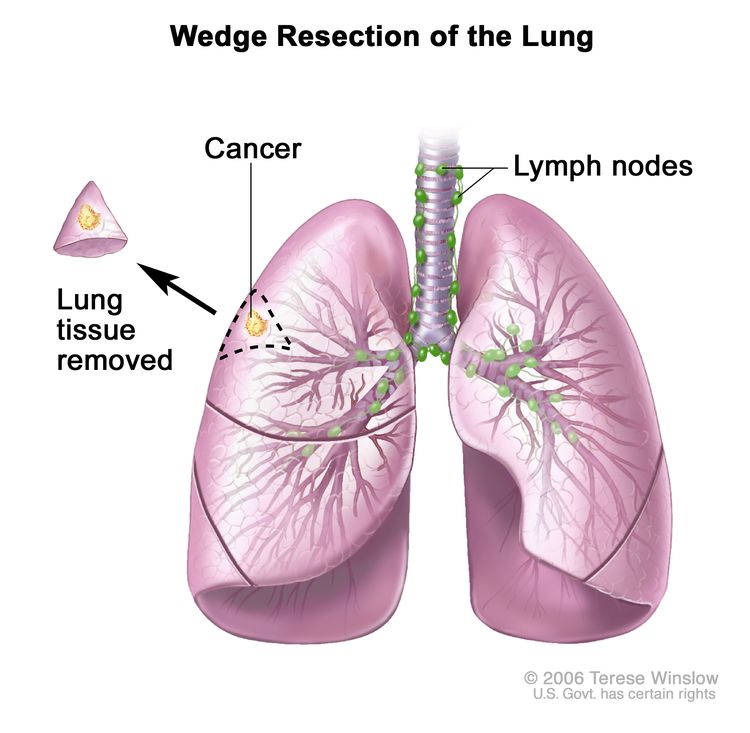 Wedge resection of the lung. Part of the lung lobe containing the cancer and a small amount of healthy tissue around it is removed.
 - Lobectomy:
Surgery to remove a whole lobe (section) of the lung.
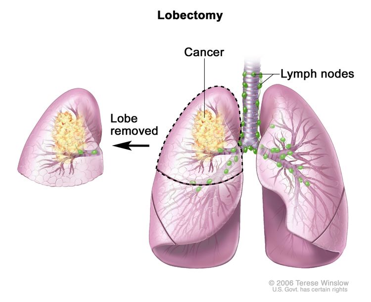 Lobectomy. A lobe of the lung is removed.
 - Pneumonectomy:
Surgery to remove one whole lung.
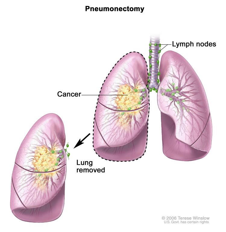 Pneumonectomy. The whole lung is removed.
 - Sleeve resection: Surgery to remove part of the bronchus.
After the doctor removes all the cancer that can be seen at the time of the surgery, some patients may be given chemotherapy or radiation therapy after surgery to kill any cancer cells that are left. Treatment given after the surgery, to lower the risk that the cancer will come back, is called adjuvant therapy.
Radiation therapy
Radiation therapy is a cancer treatment that uses high-energy x-rays or other types of radiation to kill cancer cells or keep them from growing. There are two types of radiation therapy:
- External radiation therapy uses a machine outside the body to send radiation toward the area of the body with cancer.
- Internal radiation therapy uses a radioactive substance sealed in needles, seeds, wires, or catheters that are placed directly into or near the cancer.
Stereotactic body radiation therapy is a type of external radiation therapy. Special equipment is used to place the patient in the same position for each radiation treatment. Once a day for several days, a radiation machine aims a larger than usual dose of radiation directly at the tumor. By having the patient in the same position for each treatment, there is less damage to nearby healthy tissue. This procedure is also called stereotactic external-beam radiation therapy and stereotaxic radiation therapy.
Stereotactic radiosurgery is a type of external radiation therapy used to treat lung cancer that has spread to the brain. A rigid head frame is attached to the skull to keep the head still during the radiation treatment. A machine aims a single large dose of radiation directly at the tumor in the brain. This procedure does not involve surgery. It is also called stereotaxic radiosurgery, radiosurgery, and radiation surgery.
For tumors in the airways, radiation is given directly to the tumor through an endoscope.
The way the radiation therapy is given depends on the type and stage of the cancer being treated. It also depends on where the cancer is found. External and internal radiation therapy are used to treat non-small cell lung cancer.
Chemotherapy
Chemotherapy is a cancer treatment that uses drugs to stop the growth of cancer cells, either by killing the cells or by stopping them from dividing. When chemotherapy is taken by mouth or injected into a vein or muscle, the drugs enter the bloodstream and can reach cancer cells throughout the body (systemic chemotherapy). When chemotherapy is placed directly into the cerebrospinal fluid, an organ, or a body cavity such as the abdomen, the drugs mainly affect cancer cells in those areas (regional chemotherapy).
The way the chemotherapy is given depends on the type and stage of the cancer being treated.
For more information, see Drugs Approved for Non-Small Cell Lung Cancer.
Targeted therapy
Targeted therapy is a type of treatment that uses drugs or other substances to identify and attack specific cancer cells. Monoclonal antibodies, tyrosine kinase inhibitors, mammalian target of rapamycin (mTOR) inhibitors, and KRAS G12C inhibitors are four types of targeted therapy being used to treat advanced, metastatic, or recurrent non-small cell lung cancer.
Monoclonal antibodies
Monoclonal antibodies are immune systemproteins made in the laboratory to treat many diseases, including cancer. As a cancer treatment, these antibodies can attach to a specific target on cancer cells or other cells that may help cancer cells grow. The antibodies are able to then kill the cancer cells, block their growth, or keep them from spreading. Monoclonal antibodies are given by infusion. They may be used alone or to carry drugs, toxins, or radioactive material directly to cancer cells.
There are different types of monoclonal antibody therapy:
- Vascular endothelial growth factor (VEGF) inhibitor therapy: Cancer cells make a substance called VEGF, which causes new blood vessels to form (angiogenesis) and helps the cancer grow. VEGF inhibitors block VEGF and stop new blood vessels from forming. This may kill cancer cells because they need new blood vessels to grow. Bevacizumab and ramucirumab are VEGF inhibitors and angiogenesis inhibitors.
- Epidermal growth factor receptor (EGFR) inhibitor therapy: EGFRs are proteins found on the surface of certain cells, including cancer cells. Epidermal growth factor attaches to the EGFR on the surface of the cell and causes the cells to grow and divide. EGFR inhibitors block the receptor and stop the epidermal growth factor from attaching to the cancer cell. This stops the cancer cell from growing and dividing. Cetuximab and necitumumab are EGFR inhibitors.
Tyrosine kinase inhibitors
Tyrosine kinase inhibitors are small-molecule drugs that go through the cell membrane and work inside cancer cells to block signals that cancer cells need to grow and divide. Some tyrosine kinase inhibitors also have angiogenesis inhibitor effects.
There are different types of tyrosine kinase inhibitors:
- Epidermal growth factor receptor (EGFR) tyrosine kinase inhibitors: EGFRs are proteins found on the surface and inside certain cells, including cancer cells. Epidermal growth factor attaches to the EGFR inside the cell and sends signals to the tyrosine kinase area of the cell, which tells the cell to grow and divide. EGFR tyrosine kinase inhibitors stop these signals and stop the cancer cell from growing and dividing. Erlotinib, gefitinib, afatinib, osimertinib, dacomitinib, and amivantamab are types of EGFR tyrosine kinase inhibitors. Some of these drugs work better when there is also a mutation (change) in the EGFR gene.
- Kinase inhibitors that affect cells with certain gene changes:
- Dabrafenib is used to stop proteins being made by the BRAF gene.
- Trametinib is used to stop proteins being made by the MEK gene.
- Crizotinib and entrectinib are used to stop proteins from being made by the ALK and ROS1 genes.
- Ceritinib, alectinib, brigatinib, and lorlatinib are used to stop proteins from being made by the ALK gene.
- Larotrectinib and entrectinib are used to stop proteins being made by NTRK1, NTRK2, and NTRK3 gene fusions.
- Selpercatinib and pralsetinib are used to stop proteins being made by the RET fusion gene or the mutated RET gene.
- Tepotinib and capmatinib are used to stop proteins being made by the mutated MET gene.
Mammalian target of rapamycin (mTOR) inhibitors
mTOR inhibitors block a protein called mTOR, which may keep cancer cells from growing and prevent the growth of new blood vessels that tumors need to grow. Everolimus is a type of mTOR inhibitor.
KRAS G12C inhibitors
KRAS G12C inhibitors block a protein called KRAS p.G12C, which may help keep cancer cells from growing and may kill them. Sotorasib and adagrasib are types of KRAS G12C inhibitors.
For more information, see Drugs Approved for Non-Small Cell Lung Cancer.
Immunotherapy
Immunotherapy is a treatment that uses the patient's immune system to fight cancer. Substances made by the body or made in a laboratory are used to boost, direct, or restore the body's natural defenses against cancer.
Immune checkpoint inhibitor therapy is a type of immunotherapy used to treat some patients with advanced non-small cell lung cancer.
Types of immune checkpoint inhibitor therapy include:
- PD-1 and PD-L1 inhibitor therapy: PD-1 is a protein on the surface of T cells that helps keep the body's immune responses in check. PD-L1 is a protein found on some types of cancer cells. When PD-1 attaches to PD-L1, it stops the T cell from killing the cancer cell. PD-1 and PD-L1 inhibitors keep PD-1 and PD-L1 proteins from attaching to each other. This allows the T cells to kill cancer cells. Pembrolizumab, nivolumab, and cemiplimab are types of PD-1 inhibitors. Atezolizumab and durvalumab are types of PD-L1 inhibitors.
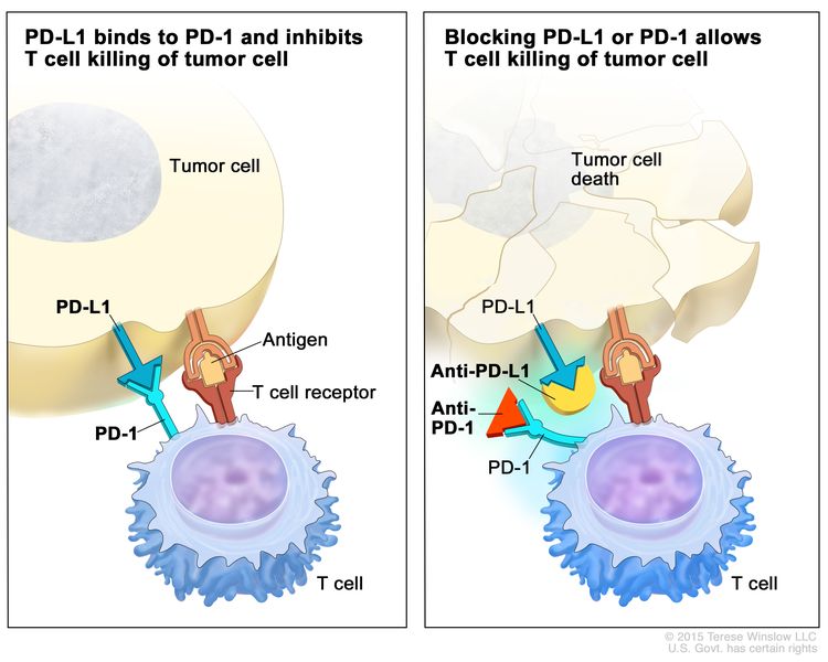 Immune checkpoint inhibitor. Checkpoint proteins, such as PD-L1 on tumor cells and PD-1 on T cells, help keep immune responses in check. The binding of PD-L1 to PD-1 keeps T cells from killing tumor cells in the body (left panel). Blocking the binding of PD-L1 to PD-1 with an immune checkpoint inhibitor (anti-PD-L1 or anti-PD-1) allows the T cells to kill tumor cells (right panel).
 - CTLA-4 inhibitor therapy: CTLA-4 is a protein on the surface of T cells that helps keep the body's immune responses in check. When CTLA-4 attaches to another protein called B7 on a cancer cell, it stops the T cell from killing the cancer cell. CTLA-4 inhibitors attach to CTLA-4 and allow the T cells to kill cancer cells. Tremelimumab is a type of CTLA-4 inhibitor.
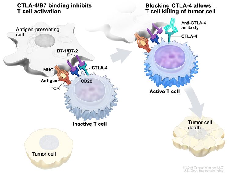 Immune checkpoint inhibitor. Checkpoint proteins, such as B7-1/B7-2 on antigen-presenting cells (APC) and CTLA-4 on T cells, help keep the body’s immune responses in check. When the T-cell receptor (TCR) binds to antigen and major histocompatibility complex (MHC) proteins on the APC and CD28 binds to B7-1/B7-2 on the APC, the T cell can be activated. However, the binding of B7-1/B7-2 to CTLA-4 keeps the T cells in the inactive state so they are not able to kill tumor cells in the body (left panel). Blocking the binding of B7-1/B7-2 to CTLA-4 with an immune checkpoint inhibitor (anti-CTLA-4 antibody) allows the T cells to be active and to kill tumor cells (right panel).
For more information, see Drugs Approved for Non-Small Cell Lung Cancer.
Laser therapy
Laser therapy is a cancer treatment that uses a laser beam (a narrow beam of intense light) to kill cancer cells.
Photodynamic therapy (PDT)
Photodynamic therapy (PDT) is a cancer treatment that uses a drug and a certain type of laser light to kill cancer cells. A drug that is not active until it is exposed to light is injected into a vein. The drug collects more in cancer cells than in normal cells. Fiberoptic tubes are then used to carry the laser light to the cancer cells, where the drug becomes active and kills the cells. Photodynamic therapy causes little damage to healthy tissue. It is used mainly to treat tumors on or just under the skin or in the lining of internal organs. When the tumor is in the airways, PDT is given directly to the tumor through an endoscope.
Cryosurgery
Cryosurgery is a treatment that uses an instrument to freeze and destroy abnormal tissue, such as carcinoma in situ. This type of treatment is also called cryotherapy. For tumors in the airways, cryosurgery is done through an endoscope.
Electrocautery
Electrocautery is a treatment that uses a probe or needle heated by an electric current to destroy abnormal tissue. For tumors in the airways, electrocautery is done through an endoscope.
New types of treatment are being tested in clinical trials.
This summary section describes treatments that are being studied in clinical trials. It may not mention every new treatment being studied. Information about clinical trials is available from the NCI website.
Radiosensitizers
Radiosensitizers are substances that make tumor cells easier to kill with radiation therapy. The combination of chemotherapy and radiation therapy given with a radiosensitizer is being studied in the treatment of non-small cell lung cancer.
New combinations
New combinations of treatments are being studied in clinical trials.
Treatment for non-small cell lung cancer may cause side effects.
For information about side effects caused by treatment for cancer, see our Side Effects page.
Patients may want to think about taking part in a clinical trial.
For some patients, taking part in a clinical trial may be the best treatment choice. Clinical trials are part of the cancer research process. Clinical trials are done to find out if new cancer treatments are safe and effective or better than the standard treatment.
Many of today's standard treatments for cancer are based on earlier clinical trials. Patients who take part in a clinical trial may receive the standard treatment or be among the first to receive a new treatment.
Patients who take part in clinical trials also help improve the way cancer will be treated in the future. Even when clinical trials do not lead to effective new treatments, they often answer important questions and help move research forward.
Patients can enter clinical trials before, during, or after starting their cancer treatment.
Some clinical trials only include patients who have not yet received treatment. Other trials test treatments for patients whose cancer has not gotten better. There are also clinical trials that test new ways to stop cancer from recurring (coming back) or reduce the side effects of cancer treatment.
Clinical trials are taking place in many parts of the country. Information about clinical trials supported by NCI can be found on NCI’s clinical trials search webpage. Clinical trials supported by other organizations can be found on the ClinicalTrials.gov website.
Follow-up tests may be needed.
Some of the tests that were done to diagnose the cancer or to find out the stage of the cancer may be repeated. Some tests will be repeated in order to see how well the treatment is working. Decisions about whether to continue, change, or stop treatment may be based on the results of these tests.
Some of the tests will continue to be done from time to time after treatment has ended. The results of these tests can show if your condition has changed or if the cancer has recurred (come back). These tests are sometimes called follow-up tests or check-ups.
occult non-small cell lung cancerTreatment of Occult Non-Small Cell Lung Cancer
For information about the treatments listed below, see the Treatment Option Overview section.
Treatment of occult non-small cell lung cancer depends on the stage of the disease. Occult tumors are often found at an early stage (the tumor is in the lung only) and sometimes can be cured by surgery.
Use our clinical trial search to find NCI-supported cancer clinical trials that are accepting patients. You can search for trials based on the type of cancer, the age of the patient, and where the trials are being done. General information about clinical trials is also available.
stage 0 non-small cell lung cancerTreatment of Stage 0 (Carcinoma in Situ)
For information about the treatments listed below, see the Treatment Option Overview section.
Treatment of stage 0 may include the following:
- Surgery (wedge resection or segmental resection).
- Photodynamic therapy, electrocautery, cryosurgery, or laser surgery for tumors in or near the bronchus.
Use our clinical trial search to find NCI-supported cancer clinical trials that are accepting patients. You can search for trials based on the type of cancer, the age of the patient, and where the trials are being done. General information about clinical trials is also available.
stage I non-small cell lung cancerTreatment of Stage I Non-Small Cell Lung Cancer
For information about the treatments listed below, see the Treatment Option Overview section.
Treatment of stage IA non-small cell lung cancer and stage IB non-small cell lung cancer may include the following:
- Surgery (wedge resection, segmental resection, sleeve resection, or lobectomy).
- Surgery followed by targeted therapy with an EGFR tyrosine kinase inhibitor, such as osimertinib.
- External radiation therapy, including stereotactic body radiation therapy for patients who cannot have surgery or choose not to have surgery.
- A clinical trial of chemotherapy or radiation therapy following surgery.
Use our clinical trial search to find NCI-supported cancer clinical trials that are accepting patients. You can search for trials based on the type of cancer, the age of the patient, and where the trials are being done. General information about clinical trials is also available.
stage II non-small cell lung cancerTreatment of Stage II Non-Small Cell Lung Cancer
For information about the treatments listed below, see the Treatment Option Overview section.
Treatment of stage IIA non-small cell lung cancer and stage IIB non-small cell lung cancer may include the following:
- Surgery (wedge resection, segmental resection, sleeve resection, lobectomy, or pneumonectomy).
- Surgery followed by chemotherapy.
- Surgery followed by targeted therapy with an EGFR tyrosine kinase inhibitor, such as osimertinib.
- Surgery followed by immunotherapy, such as atezolizumab.
- Surgery followed by radiation therapy.
- Chemotherapy followed by surgery.
- Immunotherapy, such as nivolumab, and chemotherapy followed by surgery.
- External radiation therapy for patients who cannot have surgery.
- A clinical trial of radiation therapy following surgery.
Use our clinical trial search to find NCI-supported cancer clinical trials that are accepting patients. You can search for trials based on the type of cancer, the age of the patient, and where the trials are being done. General information about clinical trials is also available.
stage III non-small cell lung cancerTreatment of Stage IIIA Non-Small Cell Lung Cancer
For information about the treatments listed below, see the Treatment Option Overview section.
Treatment of stage IIIA non-small cell lung cancer that can be removed with surgery may include the following:
- Chemotherapy followed by surgery.
- Chemotherapy and radiation therapy followed by surgery.
- Immunotherapy, such as nivolumab, and chemotherapy followed by surgery.
- Surgery followed by chemotherapy.
- Surgery followed by targeted therapy with an EGFR tyrosine kinase inhibitor, such as osimertinib.
- Surgery followed by immunotherapy, such as atezolizumab.
- Surgery followed by chemotherapy combined with radiation therapy.
- Surgery followed by radiation therapy.
- A clinical trial of new combinations of treatments.
Treatment of stage IIIA non-small cell lung cancer that cannot be removed with surgery may include the following:
- Chemotherapy and radiation therapy.
- Chemotherapy and radiation therapy followed by immunotherapy with an immune checkpoint inhibitor, such as durvalumab.
- External radiation therapy alone.
- Internal radiation therapy or laser surgery, as palliative treatment to relieve symptoms and improve the quality of life.
For more information about supportive care for signs and symptoms including cough, shortness of breath, and chest pain, see Cardiopulmonary Syndromes.
Non-small cell lung cancer of the superior sulcus, often called Pancoast tumor, begins in the upper part of the lung and spreads to nearby tissues such as the chest wall, large blood vessels, and spine. Treatment of Pancoast tumors may include the following:
- Surgery.
- Chemotherapy and radiation therapy followed by surgery.
- Radiation therapy alone.
- A clinical trial of new combinations of treatments.
Some stage IIIA non-small cell lung tumors that have grown into the chest wall may be completely removed. Treatment of chest wall tumors may include the following:
- Surgery.
- Surgery and radiation therapy.
- Radiation therapy alone.
- Chemotherapy combined with radiation therapy and/or surgery.
- A clinical trial of new combinations of treatments.
Use our clinical trial search to find NCI-supported cancer clinical trials that are accepting patients. You can search for trials based on the type of cancer, the age of the patient, and where the trials are being done. General information about clinical trials is also available.
stage III non-small cell lung cancerTreatment of Stage IIIB and Stage IIIC Non-Small Cell Lung Cancer
For information about the treatments listed below, see the Treatment Option Overview section.
Treatment of stage IIIB non-small cell lung cancer and stage IIIC non-small cell lung cancer may include the following:
- Chemotherapy followed by external radiation therapy.
- Chemotherapy and radiation therapy given over the same period of time. The radiation therapy dose may increase over time.
- Chemotherapy and radiation therapy given over the same period of time. Immunotherapy with an immune checkpoint inhibitor, such as durvalumab, may be given before or after these treatments.
- External radiation therapy alone for patients who cannot be treated with chemotherapy.
- External radiation therapy as palliative therapy, to relieve symptoms and improve the quality of life.
- Laser therapy and/or internal radiation therapy to relieve symptoms and improve the quality of life.
- Clinical trials of new external radiation therapy schedules and new types of treatment.
- A clinical trial of chemotherapy and radiation therapy combined with a radiosensitizer.
- Clinical trials of targeted therapy combined with chemotherapy and radiation therapy.
For more information about supportive care for signs and symptoms such as cough, shortness of breath, and chest pain, see the following PDQ summaries:
- Cardiopulmonary Syndromes
- Cancer Pain
Use our clinical trial search to find NCI-supported cancer clinical trials that are accepting patients. You can search for trials based on the type of cancer, the age of the patient, and where the trials are being done. General information about clinical trials is also available.
stage IV non-small cell lung cancerrecurrent/relapsed non-small cell lung cancerTreatment of Newly Diagnosed Stage IV, Relapsed, and Recurrent Non-Small Cell Lung Cancer
For information about the treatments listed below, see the Treatment Option Overview section.
Treatment of newly diagnosedstage IV, relapsed, and recurrentnon-small cell lung cancer may include the following:
- Combination chemotherapy.
- Combination chemotherapy and targeted therapy with a monoclonal antibody, such as bevacizumab, cetuximab, or necitumumab.
- Combination chemotherapy followed by more chemotherapy as maintenance therapy to help keep cancer from progressing.
- Targeted therapy with an EGFR tyrosine kinase inhibitor, such as osimertinib, dacomitinib, gefitinib, erlotinib, or afatinib.
- Targeted therapy with an ALK inhibitor, such as alectinib, lorlatinib, crizotinib, ceritinib, or brigatinib.
- Targeted therapy with a BRAF inhibitor or MEK inhibitor, such as dabrafenib or trametinib.
- Targeted therapy with crizotinib and entrectinib to stop proteins from being made by the ALK and ROS1 genes.
- Targeted therapy with a NTRK inhibitor, such as larotrectinib or entrectinib.
- Targeted therapy with a RET inhibitor, such as selpercatinib or pralsetinib.
- Targeted therapy with a MET inhibitor, such as tepotinib or capmatinib.
- Immunotherapy with an immune checkpoint inhibitor, such as pembrolizumab, cemiplimab, tremelimumab, or atezolizumab, with or without chemotherapy.
- Targeted therapy with an mTOR inhibitor, such as everolimus.
- Laser therapy and/or internal radiation therapy for tumors that are blocking the airways.
- External radiation therapy as palliative therapy, to relieve symptoms and improve the quality of life.
- Surgery to remove a second primary tumor.
- Surgery to remove cancer that has spread to the brain, followed by radiation therapy to the whole brain.
- Stereotactic radiosurgery for tumors that have spread to the brain and cannot be treated with surgery.
- A clinical trial of new drugs and combinations of treatments.
Use our clinical trial search to find NCI-supported cancer clinical trials that are accepting patients. You can search for trials based on the type of cancer, the age of the patient, and where the trials are being done. General information about clinical trials is also available.
stage IV non-small cell lung cancerrecurrent/relapsed non-small cell lung cancerTreatment of Progressive Stage IV, Relapsed, and Recurrent Non-Small Cell Lung Cancer
For information about the treatments listed below, see the Treatment Option Overview section.
Treatment of progressivestage IV, relapsed, and recurrentnon-small cell lung cancer may include the following:
- Chemotherapy.
- Targeted therapy with an EGFR tyrosine kinase inhibitor, such as erlotinib, gefitinib, afatinib, osimertinib, or amivantamab.
- Targeted therapy with an ALK inhibitor, such as crizotinib, ceritinib, alectinib, brigatinib, or lorlatinib.
- Targeted therapy with a BRAF inhibitor or MEK inhibitor, such as dabrafenib or trametinib.
- Targeted therapy with crizotinib and entrectinib to stop proteins from being made by the ALK and ROS1 genes.
- Targeted therapy with a NTRK inhibitor, such as larotrectinib or entrectinib.
- Targeted therapy with a RET inhibitor, such as selpercatinib or pralsetinib.
- Targeted therapy with a MET inhibitor, such as tepotinib or capmatinib.
- Targeted therapy with a KRAS G12C inhibitor, such as sotorasib or adagrasib.
- Targeted therapy with trastuzumab deruxtecan for HER2-positive cancers.
- Immunotherapy with an immune checkpoint inhibitor, such as nivolumab, pembrolizumab, or atezolizumab.
- Targeted therapy with an mTOR inhibitor, such as everolimus.
- A clinical trial of new drugs and combinations of treatments.
Use our clinical trial search to find NCI-supported cancer clinical trials that are accepting patients. You can search for trials based on the type of cancer, the age of the patient, and where the trials are being done. General information about clinical trials is also available.
To Learn More About Non-Small Cell Lung Cancer
For more information from the National Cancer Institute about non-small cell lung cancer, see the following:
- Lung Cancer Home Page
- Lung Cancer Prevention
- Lung Cancer Screening
- Drugs Approved for Non-Small Cell Lung Cancer
- Targeted Therapy to Treat Cancer
- Immunotherapy to Treat Cancer
- Lasers to Treat Cancer
- Photodynamic Therapy for Cancer
- Cryosurgery to Treat Cancer
- Tobacco (includes help with quitting)
- Secondhand Smoke and Cancer
For general cancer information and other resources from the National Cancer Institute, see the following:
- About Cancer
- Staging
- Chemotherapy and You: Support for People With Cancer
- Radiation Therapy and You: Support for People With Cancer
- Coping with Cancer
- Questions to Ask Your Doctor about Cancer
- For Survivors and Caregivers
About This PDQ Summary
About PDQ
Physician Data Query (PDQ) is the National Cancer Institute's (NCI's) comprehensive cancer information database. The PDQ database contains summaries of the latest published information on cancer prevention, detection, genetics, treatment, supportive care, and complementary and alternative medicine. Most summaries come in two versions. The health professional versions have detailed information written in technical language. The patient versions are written in easy-to-understand, nontechnical language. Both versions have cancer information that is accurate and up to date and most versions are also available in Spanish.
PDQ is a service of the NCI. The NCI is part of the National Institutes of Health (NIH). NIH is the federal government’s center of biomedical research. The PDQ summaries are based on an independent review of the medical literature. They are not policy statements of the NCI or the NIH.
Purpose of This Summary
This PDQ cancer information summary has current information about the treatment of non-small cell lung cancer. It is meant to inform and help patients, families, and caregivers. It does not give formal guidelines or recommendations for making decisions about health care.
Reviewers and Updates
Editorial Boards write the PDQ cancer information summaries and keep them up to date. These Boards are made up of experts in cancer treatment and other specialties related to cancer. The summaries are reviewed regularly and changes are made when there is new information. The date on each summary ("Updated") is the date of the most recent change.
The information in this patient summary was taken from the health professional version, which is reviewed regularly and updated as needed, by the PDQ Adult Treatment Editorial Board.
Clinical Trial Information
A clinical trial is a study to answer a scientific question, such as whether one treatment is better than another. Trials are based on past studies and what has been learned in the laboratory. Each trial answers certain scientific questions in order to find new and better ways to help cancer patients. During treatment clinical trials, information is collected about the effects of a new treatment and how well it works. If a clinical trial shows that a new treatment is better than one currently being used, the new treatment may become "standard." Patients may want to think about taking part in a clinical trial. Some clinical trials are open only to patients who have not started treatment.
Clinical trials can be found online at NCI's website. For more information, call the Cancer Information Service (CIS), NCI's contact center, at 1-800-4-CANCER (1-800-422-6237).
Permission to Use This Summary
PDQ is a registered trademark. The content of PDQ documents can be used freely as text. It cannot be identified as an NCI PDQ cancer information summary unless the whole summary is shown and it is updated regularly. However, a user would be allowed to write a sentence such as “NCI’s PDQ cancer information summary about breast cancer prevention states the risks in the following way: [include excerpt from the summary].”
The best way to cite this PDQ summary is:
PDQ® Adult Treatment Editorial Board. PDQ Non-Small Cell Lung Cancer Treatment. Bethesda, MD: National Cancer Institute. Updated
Images in this summary are used with permission of the author(s), artist, and/or publisher for use in the PDQ summaries only. If you want to use an image from a PDQ summary and you are not using the whole summary, you must get permission from the owner. It cannot be given by the National Cancer Institute. Information about using the images in this summary, along with many other images related to cancer can be found in Visuals Online. Visuals Online is a collection of more than 3,000 scientific images.
Disclaimer
The information in these summaries should not be used to make decisions about insurance reimbursement. More information on insurance coverage is available on Cancer.gov on the Managing Cancer Care page.
Contact Us
More information about contacting us or receiving help with the Cancer.gov website can be found on our Contact Us for Help page. Questions can also be submitted to Cancer.gov through the website’s E-mail Us.