Small Cell Lung Cancer: Staging and Treatment
What is staging for cancer?
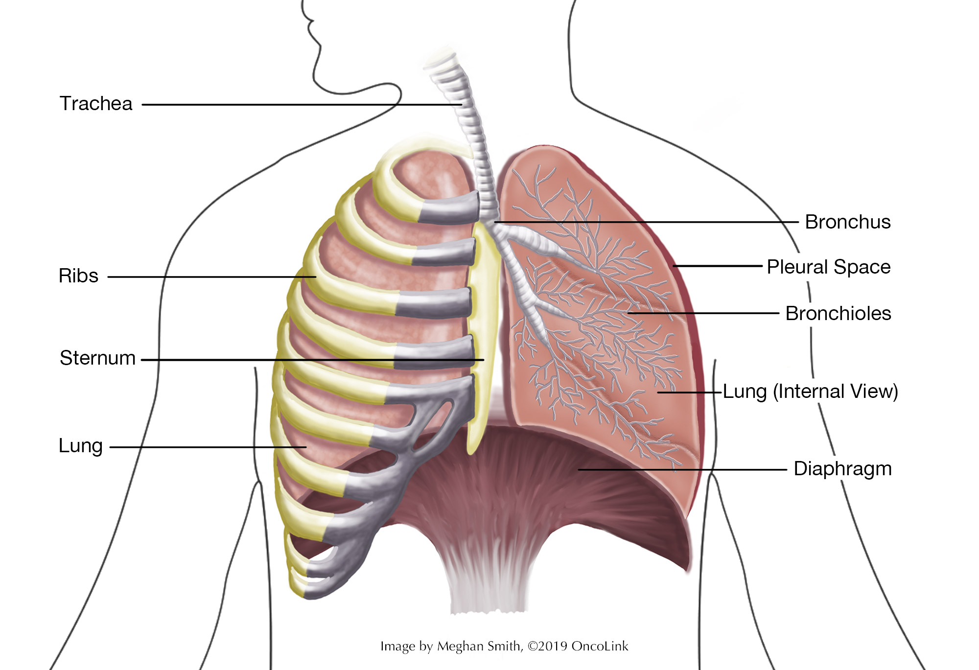
Anatomy of the chest.
Staging is the process of learning how much cancer is in your body and where it is. For small cell lung cancer (SCLC), a bronchoscopy, biopsy, chest x-ray, CT scan, MRI of the brain, and/or PET scan may be used to stage your cancer. Your providers need to know about your cancer and your health so that they can plan the best treatment for you.
How is small-cell lung cancer staged?
Staging helps guide your treatment options. Staging looks at the size of the tumor, where it is, and if it has spread to other organs.
A staging system called the “TNM system,” as described by the American Joint Committee on Cancer, helps to guide the staging of SCLC. It has three parts:
- T- Describes the size/location/extent of the "primary" tumor in the lung.
- N- Describes if the cancer has spread to the lymph nodes.
- M- Describes if the cancer has spread to other organs (metastases).
Your healthcare provider will use the results of the tests you had to determine your TNM result and combine these to get a stage from 0 (zero) to IV (four).
SCLC is often staged in two categories: limited stage or extensive stage. These stages are based on the “TNM” system.
- Limited stage (TNM Stage I-III: TI-III, any N, No M): The cancer is on one side of the chest. The cancer may have spread to the lymph nodes. The cancer has not spread to other organs
- Extensive stage (TNM stage IV: Any T, Any N, M1a/b/c): The cancer has spread in the lung, spread to the other lung, to the lymph nodes, and to other parts of the body.
The staging systems are very complex. Below is a summary. Talk to your provider about the stage of your cancer.
- Stage 0 (Tis, N0, M0): The cancer is only in the top layers of the airway and has not spread deeper into the lung (Tis). It has not spread to the lymph nodes (N0) or other parts of the body (M0).
- Stage Ia1 (T1a/mi, N0, M0): The tumor is a minimally invasive adenocarcinoma. The tumor is not larger than 3 centimeters (cm) and the part that has invaded into deeper lung tissues is not larger than ½ cm (T1mi). The cancer has not spread to nearby lymph nodes (N0) or to distant parts of the body (M0); OR (T1a, N0, M0) The tumor is no bigger than 1 cm in size. It hasn’t spread to the area around the lungs (pleura) and it hasn’t grown into the main parts of the bronchi (T1a). It has not spread to the lymph nodes (N0) or other parts of the body (M0).
- Stage IA2 (T1b, N0, M0): The tumor is between 1 and 2 cm in size. It hasn’t spread to the area around the lungs (pleura) and it hasn’t grown into the main parts of the bronchi (T1b). It has not spread to the lymph nodes (N0) or other areas of the body (M0).
- Stage IA3 (T1c, N0, M0): The tumor is 2 to 3 cm in size. It hasn’t spread to the area around the lungs (pleura) and it hasn’t grown into the main parts of the bronchi (T1c). It has not spread to the lymph nodes (N0) or other areas of the body (M0).
- Stage IB (T2a, N0, M0): The tumor is 1 or more of the following (T2a): (1) The tumor is 3 to 4 cm in size, (2) it has grown into a main bronchus but not the carina (where the windpipe splits into the left and right bronchi) and is less that 4 cm in size, (3) it has grown into the area around the lungs (pleura) and is less than 4 cm in size, or (4) it is partly blocking the airways and is less than 4 cm in size.
It has not spread to the lymph nodes (N0) or other areas of the body (M0). - Stage IIA (T2b, N0, M0): The tumor is 1 or more of the following (T2b): (1) it is 4 to 5 cm in size, (2) it has grown into the main bronchus but not the carina (where the windpipe splits into the left and right bronchi) and is 4 to 5 cm in size, (3) the tumor has grown into the area around the lungs (pleura) and is 4 to 5 cm in size, or (4) it is partly blocking the airways and is less than 5 cm in size.
It has not spread to the lymph nodes (N0) or other areas of the body (M0). - Stage IIB
- (T1a/T1b/T1c, N1, M0): The tumor is 3 cm or less in size. It has not grown into the area around the lungs (pleura) and does not affect the main branches of the bronchi (T1). It has spread to lymph nodes within the lung or in the area where the bronchus enters the lung.
These lymph nodes are on the same side as the tumor (N1). The cancer has not spread to other parts of the body (M0). - (T2a/T2b/ N1, M0): The tumor is 1 or more of the following: (1) It is 3 to 5 cm in size, (2) It has grown into a main bronchus but not the carina (where the windpipe splits into the left and right bronchi) and is less than 5 cm in size, (3) it has grown into the area around the lungs (pleura) and is less than 5 cm in size, or (4) it is partly blocking the airways and is less than 5 cm in size. It has spread to lymph nodes within the lung or in the area where the bronchus enters the lung.
These lymph nodes are on the same side as the tumor (N1). The cancer has not spread to other parts of the body (M0). - (T3, N0, M0): The tumor is one or more of the following: (1) The tumor is 5 to 7 cm in size, (2) it has grown into the chest wall, the inner lining of the chest wall, the phrenic nerve, or the area around the heart (parietal pericardium), or (3) there are 2 or more separate tumor nodules in the same lobe of a lung. It has not spread to the lymph nodes (N0) or other areas of the body (M0).
- (T1a/T1b/T1c, N1, M0): The tumor is 3 cm or less in size. It has not grown into the area around the lungs (pleura) and does not affect the main branches of the bronchi (T1). It has spread to lymph nodes within the lung or in the area where the bronchus enters the lung.
- Stage IIIA
- (T1a, T1b, T1c, N2, M0): The tumor is no bigger than 3 cm in size. It hasn’t grown into the pleura and does affect the main parts of the bronchi (T1). The cancer has spread to the lymph nodes below the carina (where the windpipe splits into the left and right bronchi) or in the space between the lungs (the mediastinum). These lymph nodes are on the same side as the lung with the main tumor (N2). The cancer has not spread to other areas of the body (M0).
- (T2a/T2b, N2, M0): The tumor is one of more of the following: (1) It is 3 to 5 cm in size, (2) it has grown into the main bronchus but not the carina (where the windpipe splits into the left/right bronchi) and is less than 5 cm in size, (3) it has grown into the pleura and is less than 5 cm in size, or 4) it is partly blocking the airways and is less than 5 cm in size.
The cancer has spread to the lymph nodes below the carina and the mediastinum. These lymph nodes are on the same side as the lung with the main tumor (N2). The cancer has not spread to other parts of the body (M0). - (T3, N1, M0): The tumor is one or more of the following: (1) it is 5 to 7 cm in size, (2) the tumor has grown into the chest wall, the inner lining of the chest wall (parietal pleura), the phrenic nerve, or the area around the heart (parietal pericardium), or (3) there are 2 or more separate tumors in the same part of the lung.
The tumor has also spread to the lymph nodes in the lung and around the lung. These lymph nodes are on the same side as the lung with the tumor (N1). The cancer has not spread to other areas of the body (M0). - (T4, N0 or N1, M0): The tumor is one or more of the following: (1) It is bigger than 7 cm in size, (2) the tumor has grown into the space between the lungs (the mediastinum), the heart, the blood vessels near the heart, the windpipe (trachea), the diaphragm, the esophagus, the spine, or the carina, or (3) there are 2 or more tumors in different lobes of the same lung.
The cancer may have also spread to the lymph nodes in the lung or near the bronchus. These lymph nodes are on the same side of the body as the tumor (N0 or N1). The cancer has not spread to other parts of the body (M0).
- Stage IIIB
- (T1a/T1b/T1c, N3, M0): The tumor is no bigger than 3 cm in size. It has not grown into the pleura or the bronchi’s main branches (T1). The cancer has spread to lymph nodes above the collarbone on either side of the body or to the lymph nodes near the other lung from the main tumor site (N3). It has not spread to other parts of the body (M0).
- (T2a/T2b, N3, M0): The tumor is one or more of the following (T2): The tumor is 3 to 5 cm in size, (2) it has spread into the main bronchus, but not the carina (where the windpipe splits into the left and right bronchi) and it is less than 5 cm in size, (3) it has grown into the pleura and is less than 5 cm in size, or (4) is partly blocking the airways and is less than 5 cm in size.
The cancer has spread to lymph nodes above the collarbone on either side of the body, and/or to the lymph nodes near the other lung on the other side of the body from the main tumor site (N3). It has not spread to other parts of the body (M0). - (T3, N2, M0): The tumor is one or more of the following (T3): (1) it is 5 to 7 cm in size, (2) it has grown into the chest wall, the inner lining of the chest wall (parietal pleura), the phrenic nerve, or the area around the heart (parietal pericardium), or (3) there are 2 or more separate tumors in the same lobe of the lung.
The cancer has also spread to the lymph nodes below the carina (where the windpipe splits into the left and right bronchi) or in the space between the lungs (the mediastinum). These lymph nodes are on the same side as the lung with the main tumor (N2). The cancer has not spread to other parts of the body (M0). - (T4, N2,M0): The tumor is one or more of the following (T4): (1) It is bigger than 7 cm in size, (2) it has grown into the mediastinum, the heart, the blood vessels near the heart, the windpipe (trachea), the diaphragm, the esophagus, the spine, or the carina (where the windpipe splits into the left and right bronchi), or (3) there are 2 or more tumors in different lobes of the same lung.
The cancer has also spread to the lymph nodes below the carina and the mediastinum. These lymph nodes are on the same side as the lung with the main tumor (N2). The cancer has not spread to other areas of the body (M0).
- Stage IIIC
- (T3, N3, M0): The tumor is one or more of the following (T3): (1) it is 5 to 7 cm in size, (2) it has grown into the chest wall, the inner lining of the chest wall (parietal pleura), the phrenic nerve, or the area around the heart (parietal pericardium), or (3) there are 2 or more separate tumors in the same lobe of the lung.
The cancer has spread to lymph nodes above the collarbone on either side of the body and/or to the lymph nodes near the other lung on the other side of the body from the main tumor site (N3). It has not spread to other parts of the body (M0). - (T4, N3, M0): The tumor is one or more of the following (T4): (1) It is bigger than 7 cm in size, (2) the tumor has grown into the space between the lungs (the mediastinum), the heart, the blood vessels near the heart, the windpipe (trachea), the diaphragm, the esophagus, the spine, or the carina (where the windpipe splits into the left and right bronchi), or (3) there are 2 or more tumors in different lobes of the same lung.
The cancer has spread to lymph nodes above the collarbone on either side of the body and/or to the lymph nodes near the other lung on the other side of the body from the main tumor site (N3). It has not spread to other parts of the body (M0).
- (T3, N3, M0): The tumor is one or more of the following (T3): (1) it is 5 to 7 cm in size, (2) it has grown into the chest wall, the inner lining of the chest wall (parietal pleura), the phrenic nerve, or the area around the heart (parietal pericardium), or (3) there are 2 or more separate tumors in the same lobe of the lung.
- Stage IVA
- (Any T, Any N, M1a): The tumor can be any size and may have grown into the pleura, bronchi, mediastinum, or other nearby parts of the body (Any T). It may have spread to nearby lymph nodes (Any N). It may also (M1a) (1) have spread to the other lung, (2) have spread to either the lining around the lungs (pleura) or the pericardium (lining around the heart), or (3) have caused cancer cells in the fluid around the lung (malignant pleural effusion), or (4) cancer cells in the fluid around the heart (malignant pericardial effusion).
- (Any T, Any N, M1b): The tumor can be any size and may have grown into the pleura, bronchi, mediastinum, or other nearby parts of the body (Any T). It may have spread to nearby lymph nodes (Any N). The tumor has also spread as a single tumor outside of the chest. This could be a lymph node in another part of the body, or in an organ such as the brain, liver, or bone (M1b).
- Stage IVB
- (Any T, Any M, M1c): The tumor can be any size and may have grown into the pleura, bronchi, mediastinum, or other nearby parts of the body (Any T). It may have spread to nearby lymph nodes (Any N). The tumor has also spread to more than 1 tumor outside of the chest. This could be a lymph node in another part of the body, and/or the brain, the liver, or the bone (M1c).
How is small-cell lung cancer treated?
Treatment for SCLC depends if your cancer is limited or extensive-stage SCLC. If you smoke, quit as soon as possible. Smoking may lessen how well your cancer treatments work and can make the side effects of treatment worse.
Your treatment may include:
- Surgery.
- Chemotherapy.
- Immunotherapy.
- Radiation therapy/PCI.
- Palliative treatment.
- Clinical trials.
Surgery
Surgery is not often used to treat SCLC unless your cancer is very early stage and hasn’t spread to your lymph nodes. This surgery would remove the lobe of the lung with the tumor and would be used with other treatments like chemotherapy and radiation.
Chemotherapy
Chemotherapy is the use of anti-cancer medicines that go through your whole body. These medicines may be given through a vein (IV, intravenously) or by mouth. Chemotherapy for SCLC may be used with immunotherapy and/or radiation therapy. What treatment you get and how often you have treatment will depend on if you have limited or extensive-stage cancer.
The medications used to treat SCLC include carboplatin, etoposide, cisplatin, topotecan, lurbinectedin, paclitaxel, docetaxel, irinotecan, temozolomide, cyclophosphamide, doxorubicin, vincristine, vinorelbine, gemcitabine, and bendamustine.
Your provider will talk to you about what regimen (type, dose, and schedule of your treatments) you will get and the potential side effects.
Immunotherapy
Immunotherapy medications work with the immune system to kill cancer cells. Immunotherapy medications that may be used in the treatment of SCLC are nivolumab, ipilimumab, pembrolizumab, atezolizumab and durvalumab. Your provider will talk to you about if these medications will help treat your cancer and what side effects you may experience.
Radiation and Prophylactic Cranial Irradiation (PCI)
Radiation is the use of high-energy X-rays to kill cancer cells. If you have limited-stage SCLC radiation may be given with chemotherapy or after chemotherapy. In extensive-stage SCLC radiation to the chest may be used after chemotherapy. Radiation may also be used to treat areas where your cancer has metastasized like the brain or bone.
Prophylactic cranial irradiation (PCI) can lower the risk of the cancer spreading to the brain. This can be done in patients with limited or extensive-stage SCLC.
Palliative Treatment
Palliative therapies are used to relieve symptoms that are caused by the cancer. They do not cure the cancer. There are many options for palliative treatments, including chemotherapy, radiation, surgery, stent placement, laser therapies, and removal of extra fluid from around the heart or lungs. Talk to your provider about your options for managing your symptoms.
Clinical Trials
You may be offered a clinical trial as part of your treatment plan. To find out more about current clinical trials, visit the OncoLink Clinical Trials Matching Service.
Making Treatment Decisions
Your care team will make sure you are a part of choosing your treatment plan. This can be overwhelming as you may be given a few options to choose from. Take the time to meet with different providers and think about your options and what is best for you. This is a personal decision. Friends and family can help you talk through the options and the pros and cons of each, but they cannot make the decision for you. You need to be comfortable with your decision – this will help you move on to the next steps. If you ever have any questions or concerns, be sure to call your team.