All About Ovarian Cancer

Author: Marisa Healy, BSN, RN
Content Contributor: Christopher Dolinsky, MD and Carolyn Vachani, MSN, RN, AOCN
Last Reviewed:

What are the ovaries?

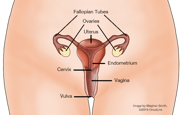
The ovaries are two small organs found only in women. They are in a woman's pelvis, connected to her uterus (the organ where a baby grows and develops when a woman is pregnant) by the fallopian tubes. The ovaries are each about the size of a marble, and they can often be felt by your healthcare provider during a pelvic examination.

The ovaries have two jobs in a woman’s body:

  1. They make female hormones.
  2. They make eggs.

Every month that a woman is fertile and not pregnant, her ovaries release a mature egg that travels into her uterus and could become fertilized. The ovaries also make estrogen and progesterone. These hormones regulate a woman's menstrual cycles, help the development during puberty, and keep a woman fertile.

What is ovarian cancer?

Ovarian cancer happens when cells in the ovaries begin to grow out of control. These cells can also invade nearby tissue or spread throughout the body. Groups of these "out-of-control" cells are often called tumors. Some tumors are not cancer because they cannot spread to other parts of the body. These are called benign tumors or masses. The tumors that can spread through the body or invade nearby tissues are true invasive cancer, also called malignant tumors. 

Many ovarian tumors are benign (not cancer). Also, women (especially younger women) can get ovarian cysts, which are collections of fluid in the ovaries that can grow large or cause pain. Ovarian cysts are not cancer. Your healthcare provider may suggest that you have an ovarian cyst removed if it is big or painful. 

Cancers are characterized by the cells from which they originally form.

  • The most common type of ovarian cancer is called epithelial ovarian cancer. This type comes from cells that lie on the surface of the ovary known as epithelial cells. Epithelial ovarian cancer makes up about 90% of all ovarian cancers and usually occurs in older women.
  • About 5% of ovarian cancers are called germ cell ovarian cancers and start in the ovarian cells that produce eggs. Germ cell ovarian cancers are more likely to affect younger women.
  • Another 5% of ovarian cancers are known as stromal ovarian cancers and develop from the cells in the ovary that hold the ovary together and make hormones. These tumors can create symptoms by making too much of the female hormones.

Each of these three types of ovarian cancer (epithelial, germ cell, stromal) has many subtypes of cancer based on how the cells look under a microscope. Talk with your provider about your exact type of ovarian cancer.

A rare type of cancer, called primary peritoneal cancer, is a malignant tumor starting in the peritoneum (the lining of the abdominal [belly] cavity). It behaves like ovarian cancer, and they can look the same under a microscope. The treatments used are often the same as those used for ovarian cancer. This type of cancer can develop in women with or without ovaries.

What causes ovarian cancer and am I at risk?

In the U.S., about 21,410 women will be diagnosed with ovarian cancer each year. Ovarian cancer is quite rare compared to other cancers.

No one knows exactly why one woman gets ovarian cancer and another does not. The most important risk factor is age. The older a woman is, the higher her chances are of ovarian cancer. The median age at diagnosis is 63, although women with genetic or family risk factors tend to be diagnosed with ovarian cancer at a younger age. 

The next risk factor for ovarian cancer is a family history of ovarian cancer. This is especially important if your family members are affected at an early age. If your mother, sister(s), or daughter(s) have had ovarian cancer, you are at an increased risk of getting ovarian cancer. It is estimated that 7% to 10% of all ovarian cancers are the result of hereditary genetic syndromes. Genetic mutations that increase ovarian cancer risk include:

It may be helpful to test for mutations if you have a strong family history of breast or ovarian cancer (multiple relatives affected, especially if they are under 50 years old when they get the disease). Having a mutation doesn't always mean you are going to be diagnosed with ovarian cancer, but it does greatly increase your chances above the general population. If you do have a mutation, you can get more rigorous screening, take preventive medications, or have prophylactic oophorectomies (preventive removal of your ovaries) to decrease your cancer risk. The decision to have genetic testing is a highly personal one. You should talk with your healthcare provider and a genetic counselor who is trained in counseling patients about genetic testing and results.

Other risk factors include being overweight, never having children or having them later in life, using fertility treatments, taking hormone therapy after menopause, smoking, and alcohol use. Keep in mind someone without any risk factors can still get ovarian cancer. Talk to your healthcare provider about your risk factors for ovarian cancer to their recommendations for screening and prevention.

How can I prevent ovarian cancer?

If you are a woman without a family history/genetic syndrome, the best way to prevent ovarian cancer is to change whatever risk factors you have control over. For example, having children by age 30 and breastfeeding both reduce risk. The use of oral contraceptives for 4 or more years is linked with lower ovarian cancer risk in the general population. Bilateral tubal ligation and hysterectomy also decrease ovarian cancer risk. Keep in mind that any medication or surgical procedure has its own risks and should be discussed with your care team.

Women who are carriers of one of the genetic syndromes listed above face different decisions. They generally need to have more rigorous screening done for ovarian cancer. They may want to take a medication, such as tamoxifen, to reduce their risk, and some may choose to have their ovaries removed when they are still healthy (called a prophylactic oophorectomy). This should only be done when a woman is finished having children. It can greatly reduce a woman's chances for getting ovarian cancer (but not reduce the risk to zero). Before a woman decides to do this, she should have genetic testing and counseling from a healthcare provider who has experience with genetic diseases.

While a diet high in animal fats has been found to increase the risk of ovarian cancer, a diet rich in fruits and vegetables may have a small preventive effect. Supplementation with vitamins A, C, and E may decrease your risk. However, there are no official nutritional recommendations that can be made to prevent ovarian cancer.

What screening tests are used for ovarian cancer?

If you are at average risk for ovarian cancer, there are no recommended screening tests. However, you should have a gynecological exam as often as your provider suggests. Your provider may be able to feel your ovaries during a pelvic exam, and if any changes are felt, you may need further tests. Early ovarian cancers aren't usually felt on examination, and are often missed.

There are a few other tests that are currently being studied for ovarian cancer screening. One is a blood test that looks for a protein named CA-125. CA-125 is a protein that is shed from damaged ovary cells and is often high in ovarian cancer. There are a few problems with CA-125 as a screening test. The level can be high in many other diseases besides ovarian cancer, including other cancers, endometriosis, fibroids, menstruation, colitis, diverticulitis, pancreatitis, lupus, and inflammation of the lining of the lung or heart. 

One possible way to use CA-125 for ovarian cancer screening is to check it and then re-check it 6 months later. If it greatly increases over this time, then ovarian cancer is more likely. The problem is that many patients without ovarian cancer will have elevated CA-125 levels and then have unneeded further workup (called a false positive). You could also have a false negative result, where the CA-125 is not higher, but there is actually cancer present and it goes unrecognized.

Another method for ovarian cancer screening is transvaginal ultrasound. Ultrasound is an imaging test that uses sound waves that bounce off of tissues and provide a picture of whatever is being looked at. By inserting an ultrasound probe into a woman's vagina, healthcare providers can get a good look at her ovaries. If the ovaries look abnormal, further tests can be done. The biggest problem with using transvaginal ultrasound for ovarian cancer screening is the same problem as using CA-125. Both tests cause too many healthy women to have unneeded procedures because the tests are not specific enough for ovarian cancer. In studies, combining CA-125 and vaginal ultrasound did not lower the number of deaths related to ovarian cancer, and therefore are not routinely recommended for screening.

Women with a strong family history or those with a proven hereditary cancer syndrome may need to get more rigorous screening with serial CA-125 tests and/or transvaginal ultrasounds, though the benefit to this is not clear. Talk to your healthcare provider about your ovarian cancer risk, and the best way to go about screening in your case.

What are the signs of ovarian cancer?

Ovarian cancer has been called a "silent killer" because it was thought that symptoms did not develop until the disease was advanced. More recently, ovarian cancer experts found that this was not true. Most women had symptoms early on that were dismissed by themselves or their healthcare providers. Experts worked together to develop the Ovarian Cancer Symptoms Consensus Statement, which describes important symptoms.

The symptoms that are more likely seen in women with ovarian cancer compared with healthy women include:

  • Bloating (feeling full).
  • Pelvic or abdominal (belly) pain.
  • Having a hard time eating or feeling full quickly.
  • Urinary changes (urgency or frequency).

While these symptoms are more often due to other medical problems, women with ovarian cancer report that the symptoms don’t go away and are a change from their normal. The frequency and number of these symptoms are also key factors in the diagnosis.

Other possible symptoms include fatigue, indigestion, back pain, painful intercourse, constipation, and menstrual changes. Women who have these symptoms almost daily for more than a few weeks should see a healthcare provider (preferably a gynecologist) for testing.

How is ovarian cancer diagnosed?

The most common reason for a healthcare provider to suspect ovarian cancer is if he/she feels a mass during a pelvic examination. Ovarian cancer is a type of cancer that needs to be diagnosed and staged during a surgery. Often, the cancer is diagnosed and treated during the same procedure. Surgeries for ovarian cancer diagnosis and treatment should be done by a surgeon who specializes in gynecologic malignancies. Surgery is done so that samples of the mass and surrounding tissue can be biopsied and analyzed. A biopsy is the only way to know for sure if you have cancer, because it allows your healthcare providers to obtain cells that can be examined under a microscope. Once the tissue is removed, a healthcare provider called a pathologist will review the specimen. The pathologist can tell if it is cancer or not; and if it is cancerous, the pathologist will characterize it by what type of tissue it arose from and what subtype of ovarian cancer it is, how abnormal it looks (called the grade), and whether or not it is invading surrounding tissues.

Although surgery is required for accurate staging, your providers may want to order some other tests to better characterize the mass/masses and look for distant spread. Tests like CT scans or MRIs can examine the pelvis and localized lymph nodes. Some patients with bony pain are referred for a bone scan, which is a test using a radioactive tracer to look for metastasis to any of the bones. You may also undergo a colonoscopy, which uses a lighted scope to examine your rectum and colon, or a barium enema in which dye is inserted into your rectum and an x-ray is taken. These tests are used to look for spread of the tumor to your colon. Each patient is unique, so the specific tests people get will vary; but overall, your providers want to know as much about your particular tumor as possible so that they can plan the best available treatments.

How is ovarian cancer staged?

In order to guide treatment, ovarian cancer is staged into four different groups. The staging system used for ovarian cancer is the FIGO system (International Federation of Gynecologists and Obstetricians). Healthcare providers also use the TNM system (also called tumor - node - metastasis system). TNM describes:

  • T: The size and local invasiveness of the tumor.
  • N: Which, if any, lymph nodes are involved.
  • M: If it has spread to other more distant areas of the body.

This is then interpreted as a stage somewhere from I (one) denoting more limited disease to IV (four) denoting more advanced disease. Generally, the higher the stage, the more serious the cancer.

In addition to stage, the tumor grade will also be evaluated. This refers to how abnormal cells appear under a microscope. Low grade (or grade I) tumors appear the most like normal cells, whereas higher grade tumors (grades 3 and 4) appear very abnormal under the microscope. Higher-grade tumors may behave more aggressively than low-grade tumors. The staging system for ovarian and primary peritoneal cancer is also applied to malignant germ cell tumors, malignant sex cord-stromal tumors, and carcinosarcoma (malignant mixed Müllerian tumors).

The staging system is very complex, and the entire staging system is outlined at the end of this article. Though complicated, the staging system helps healthcare providers determine the extent of the cancer, and in turn, make treatment decisions for a patient's cancer. The stage of cancer, or extent of disease, is based on information gathered through the various tests done as the diagnosis and work-up of the cancer is being performed. 

How is ovarian cancer treated?

Surgery

Almost all women with ovarian cancer will have some type of surgery during their treatment. The purpose of surgery is first to diagnose and stage the cancer, as well as to remove as much of the cancer as possible. In early-stage cancers (stage I and II), surgeons can often remove all of the visible cancer. Generally, women with ovarian cancer will have a hysterectomy (removal of the uterus) and bilateral salpingo-oophorectomy (removal of both ovaries and fallopian tubes) as part of their operation. This is because there is always a risk of microscopic disease in both of the ovaries and the uterus. The only time a woman may not have this entire operation is if she has a very early-stage cancer (IA) that looks favorable under the microscope (grade 1). This is often the case with germ cell ovarian tumors. If a woman's tumor has these characteristics and she wishes to keep the ability to have children, then the surgeon can remove only her diseased ovary and tube. Then, after she is done having children, she will need to have her uterus and the other tube and ovary removed. With any other stage or grade of tumor, or in patients finished with childbearing, the entire operation should be done in order to provide the best chance for a cure.

Women who have more advanced disease (stage III or IV) will often have debulking surgeries. This means that the surgeon will remove as much disease as possible. Data collected in many studies has shown that the more cancer that is removed, the better the long-term outcome for the patient.

Sometimes, a patient will have debulking surgery and then later the cancer will come back (recurrence). It may be useful to debulk the tumor a second time, particularly if it has been at least a year between the first surgery and the recurrence. In patients with very advanced ovarian cancer, surgery may be used for palliation, meaning that patient is operated on with the intent of easing their pain or symptoms, rather than trying to cure their disease.

Operations for ovarian cancer should be done by surgeons who are trained in dealing with gynecologic malignancies because there are special skills and techniques needed to deal with these tumors.

Chemotherapy

Even if the tumors are removed during surgery, there is always a risk of recurrence because there may be microscopic cancer cells left that the surgeon cannot see or remove. In order to lower the risk of recurrence, a patient may have chemotherapy. Chemotherapy is the use of anti-cancer medications that work systemically (throughout the entire body) and are given intravenously (IV, directly into a vein), directly into the abdomen (intraperitoneal, “IP”) or orally (by mouth). Most patients with ovarian cancer will be offered chemotherapy as well as surgery (adjuvant chemotherapy). The higher the stage of cancer, the more important it is that you receive chemotherapy. Generally, only very early-stage cancers (early stage I) that look favorable under the microscope (grade 1 or 2) can be treated with surgery alone. Any woman with a more advanced stage or grade ovarian cancer should be offered chemotherapy.

There are many different chemotherapies used, and treatments often combine several medications to create a regimen. For the treatment of ovarian cancers, chemotherapy is often given intravenously (into a vein) or directly into the abdomen (intraperitoneal, "IP").

Some of the chemotherapy medications used in ovarian cancer treatment include: cisplatincarboplatindoxorubicintopotecanifosfamidedoxorubicin liposomaldocetaxelpaclitaxel, altretamine, capecitabine, cyclophosphamide, etoposide, gemcitabine, irinotecan, melphalan, pemetrexed, and vinorelbine. Most regimens contain a platinum compound chemotherapy, such as cisplatin, and a taxane, such as paclitaxel or docetaxel

Hormonal therapies including aromatase inhibitors (example: letrozole)tamoxifen, and luteinizing hormone-releasing hormone (LHRH) agonists (example: leuprolide acetate), are used more commonly in ovarian stromal type tumors, but not often in epithelial ovarian cancer.

There are also some targeted agents used in the treatment of ovarian cancer including bevacizumab, rucaparib, niraparib and olaparib. Bevacizumab is used to stop blood flow to the tumor and works best when given with chemotherapy. Rucaparib, niraparib and olaparib are used only for certain subtypes of ovarian cancer associated with BRCA mutation. Your care team will test the type of tumor you have to see if targeted treatment may be an option after other first line methods of treatment have not been successful. Olaparib and rucaparib can also be used to treat advanced cancer with or without mutations in the BRCA gene. 

Intraperitoneal (IP) chemotherapy is given directly into the abdomen through a catheter, allowed to "dwell,” or remain in the abdomen for a while, coating the area in chemotherapy. This allows high doses of the chemo to be in direct contact with the cancer cells in the abdominal cavity. After several hours, fluid is drained from the abdomen, releasing the remaining chemotherapy from the abdomen. It is typically used in stage III cancers where the tumor has been optimally debulked (leaving nothing larger than 1cm). This can also be done at the time of the debulking surgery. 

There are advantages and disadvantages to each of the different regimens that your  care team will discuss with you. Based on your health, your personal values and wishes, and possible side effects, you can work with your healthcare providers to come up with the best regimen for your cancer and your lifestyle.

Radiation

Radiation therapy uses high energy rays (similar to x-rays) to kill cancer cells. It comes from an external source, and you will need to come in 5 days a week for several weeks to a radiation therapy treatment center. The treatment takes just a few minutes and is painless. Radiation therapy is sometimes used with surgery in patients who have stage II tumors with low bulk disease. Radiation can also be used to ease the pain of metastases and/or to stop tumors from bleeding. Healthcare providers try to limit the amount of radiation that your vital organs receive, and don't like to treat large parts of the bowel and pelvis for this reason. This makes radiation less useful in ovarian cancer, where disease is often spread throughout the abdomen and pelvis. A radiation therapy team can answer questions about the process and side effects of radiation therapy in your particular case.

Clinical Trials

Clinical trials are extremely important in furthering our knowledge of this disease. It is through clinical trials that we know what we do today, and many exciting new therapies are currently being tested. Talk to your healthcare provider about participating in clinical trials in your area. You can also explore currently open clinical trials using the OncoLink Clinical Trials Matching Service

Follow-up Care and Survivorship

Once you have been treated for ovarian cancer, you need to be closely followed for a recurrence. At first, you will have follow-up visits often, usually every few months. The longer you are free of disease, the less often you will have to go for checkups. Your healthcare provider will tell you when they want follow-up visits, CA-125 levels, pelvic ultrasounds and/or CT scans, depending on your case. Your healthcare provider will also perform pelvic examinations. It is very important that you let your healthcare provider know about any symptoms you are having and that you keep all of your follow-up appointments.

Fear of recurrence, relationships and sexual health, financial impact of cancer treatment, employment issues, and coping strategies are common emotional and practical issues experienced by ovarian cancer survivors. Your healthcare team can identify resources for support and management of these challenges faced during and after cancer.

Cancer survivorship is a relatively new focus of oncology care. With almost 17 million cancer survivors in the US alone, there is a need to help patients transition from active treatment to survivorship. What happens next, how do you get back to normal, what should you know and do to live healthy going forward? A survivorship care plan can be a first step in educating yourself about navigating life after cancer and helping you communicate knowledgeably with your healthcare providers. Create a survivorship care plan today on OncoLink.

Resources For More Information

National Ovarian Cancer Coalition
Through national programs and local Chapter initiatives, the NOCC's goal is to make more people aware of the early symptoms of ovarian cancer. In addition, the NOCC provides information to assist the newly diagnosed patient, to provide hope to survivors, and to support caregivers.

Ovarian Cancer National Alliance
Connects survivors, women at risk, caregivers and health providers with the information and resources they need.

Pregnant with Cancer
Dedicated to providing women diagnosed with cancer while pregnant with information, support and hope.

Foundation for Women’s Cancers
The Foundation offers comprehensive information by cancer type that can help guide you through your diagnosis and treatment. They also offer the ‘Sisterhood of Survivorship’ to connect with others facing similar challenges.

Appendix: Complete Fallopian Tube & Ovarian Cancer Staging

AJCC, Cancer Staging Manual, 8th Edition

Primary Tumor (T)

FIGO

Description

TX

Primary tumor cannot be assessed

T0

No evidence of primary tumor

T1

I

Tumor limited to the ovaries (one or both) or fallopian tubes

T1a

IA

Tumor limited to one ovary (capsule intact) or fallopian tube, no tumor on surface; no malignant cells in peritoneal washings or ascites

T1b

IB

Tumor limited to both ovaries (capsules intact) or tubes, no tumor on surface; no malignant cells in peritoneal washings or ascites

T1c

IC

Tumor limited to one or both ovaries or tubes with any of the following:  

T1c1

IC1

Surgical spill

T1c2

IC2

Capsule ruptured before surgery or tumor on ovary or tube surface

T1c3

IC3

Malignant cells in ascites or peritoneal washings

T2

II

Tumor involves one or both Fallopian tubes with pelvic extension below pelvic brim or primary peritoneal cancer

T2a

IIA

Extension and/or implants on the uterus and/or fallopian tube(s) and/or ovaries

T2b

IIB

Extension to and/or implants on other pelvic tissues

T3

III

Tumor involves one or both fallopian tubes, or primary peritoneal cancer with microscopically confirmed peritoneal metastasis outside the pelvis and/or metastasis to retroperitoneal (pelvic and/or para-aortic) lymph nodes

T3a

IIIA2

Microscopic extrapelvic (above the pelvic brim) peritoneal involvement with or without positive retroperitoneal lymph nodes

T3b

IIIB

Macroscopic peritoneal metastasis beyond pelvis 2cm or less in greatest dimension with or without positive metastasis to the retroperitoneal lymph nodes

T3c

IIIC

Macroscopic peritoneal metastasis beyond the pelvis more than 2cm in greatest dimension with or without metastasis to the retroperitoneal lymph nodes (includes extension of tumor to capsule of liver and spleen without parenchymal involvement of either organ)


Regional Lymph Nodes (N)

FIGO

Description

NX

Regional lymph nodes cannot be assessed

N0

No regional lymph node metastasis

N0(i+)


Isolated tumor cells, < 0.2mm, in regional lymph nodes

N1

IIIA1

Positive retroperitoneal lymph nodes only (histologically confirmed)

N1a

IIIA1i

Metastasis up to and including 10mm in greatest dimension

N1b

IIIA1ii

Metastasis > 10mm in greatest dimension


Distant Metastasis (M)

FIGO

Description

M0

No distant metastasis

M1

IV

Distant metastasis, including pleural effusion with positive cytology; liver or splenic parenchymal metastasis; metastasis to extra-abdominal organs (including inguinal lymph nodes and lymph nodes outside the abdominal cavity); and transmural involvement of the intestine

M1a

IVA

Pleural effusion with positive cytology

M1b

IVB

Liver or splenic parenchymal metastases; metastases to extra-abdominal organs (including inguinal lymph nodes and lymph noes outside the abdominal cavity); transmural involvement of intestine


Stage Grouping

T

N

M

Stage 1

T1

N0

M0

Stage IA

T1a

N0

M0

Stage IB

T1b

N0

M0

Stage IC

T1c

N0

M0

Stage II

T2

N0

M0

Stage IIA

T2a

N0

M0

Stage IIB

T2b

N0

M0

Stage IIIA1

T1/2

N1

M0

Stage IIIA2

T3a

Nx, N0, N1

M0

Stage IIIB

T3b

Nx, N0, N1

M0

Stage IIIC

T3c

Nx, N0, N1

M0

Stage IV

Any T

Any N

M1

Stage IVA

Any T

Any N

M1a

Stage IVB

Any T

Any N

M1b

References

SEER Stat Fact Sheets: Ovarian Cancer

NIH BRCA1 and BRCA2: Cancer Risk and Genetic Testing Fact Sheet

Foundation for Women’s Cancer Ovarian Cancer Symptoms Consensus Statement

American Cancer Society. Ovarian Cancer. 2019. https://www.cancer.org/cancer/ovarian-cancer/about/key-statistics.html

Ansaloni, L., Agnoletti, V., Amadori, A., Catena, F., Cavaliere, D., Coccolini, F., ... & Ghermandi, C. (2012). Evaluation of extensive cytoreductive surgery and hyperthermic intraperitoneal chemotherapy (HIPEC) in patients with advanced epithelial ovarian cancer. International Journal of Gynecological Cancer, 22(5), 778-785.

Baldwin, L. A., Huang, B., Miller, R. W., Tucker, T., Goodrich, S. T., Podzielinski, I., ... & Seamon, L. G. (2012). Ten-year relative survival for epithelial ovarian cancer. Obstetrics & Gynecology, 120(3), 612-618.

Berliner, J. L., Fay, A. M., Cummings, S. A., Burnett, B., & Tillmanns, T. (2013). NSGC practice guideline: risk assessment and genetic counseling for hereditary breast and ovarian cancer. Journal of Genetic Counseling, 22(2), 155-163.

Bristow, R. E., Powell, M. A., Al-Hammadi, N., Chen, L., Miller, J. P., Roland, P. Y., ... & Cliby, W. A. (2013). Disparities in ovarian cancer care quality and survival according to race and socioeconomic status. Journal of the National Cancer Institute, 105(11), 823-832.

Crum, C. P. (2015). Preventing ovarian cancer. Journal of Clinical Oncology, JCO-2015.

Dinkic, C., Eichbaum, M., Schmidt, M., Grischke, E. M., Gebauer, G., Fricke, H. C., ... & Sohn, C. (2017). Pazopanib (GW786034) and cyclophosphamide in patients with platinum-resistant, recurrent, pre-treated ovarian cancer-Results of the PACOVAR-trial. Gynecologic Oncology.

Havrilesky, L. J., Moorman, P. G., Lowery, W. J., Gierisch, J. M., Coeytaux, R. R., Urrutia, R. P., ... & Myers, E. R. (2013). Oral contraceptive pills as primary prevention for ovarian cancer: a systematic review and meta-analysis. Obstetrics & Gynecology, 122(1), 139-147.

Jayson, G. C., Kohn, E. C., Kitchener, H. C., & Ledermann, J. A. (2014). Ovarian cancer. The Lancet, 384(9951), 1376-1388.

Kaijser, J., Bourne, T., Valentin, L., Sayasneh, A., Van Holsbeke, C., Vergote, I., ... & Timmerman, D. (2013). Improving strategies for diagnosing ovarian cancer: a summary of the International Ovarian Tumor Analysis (IOTA) studies. Ultrasound in Obstetrics & Gynecology, 41(1), 9-20.

Kipps, E., Tan, D. S., & Kaye, S. B. (2013). Meeting the challenge of ascites in ovarian cancer: new avenues for therapy and research. Nature Reviews Cancer, 13(4), 273-282.

Kristeleit, R., Shapiro, G. I., Burris, H. A., Oza, A. M., LoRusso, P. M., Patel, M., ... & Safra, T. (2017). A phase I-II study of the oral poly (ADP-ribose) polymerase inhibitor rucaparib in patients with germline BRCA1/2-mutated ovarian carcinoma or other solid tumors. Clin Cancer Res23(15), 4094-4106.

Ledermann, J., Harter, P., Gourley, C., Friedlander, M., Vergote, I., Rustin, G., ... & Matei, D. (2012). Olaparib maintenance therapy in platinum-sensitive relapsed ovarian cancer. New England Journal of Medicine, 366(15), 1382-1392.

Matz, M., Coleman, M. P., Carreira, H., Salmerón, D., Chirlaque, M. D., Allemani, C., & CONCORD Working Group. (2017). Worldwide comparison of ovarian cancer survival: Histological group and stage at diagnosis (CONCORD-2). Gynecologic Oncology144(2), 396-404.

Morrison, J., Haldar, K., Kehoe, S., & Lawrie, T. A. (2012). Chemotherapy versus surgery for initial treatment in advanced ovarian epithelial cancer. Cochrane Database Syst Rev, 8, CD005343.

NCCN Clinical Practice Guidelines. 2.2019. Found at: https://www.nccn.org/professionals/physician_gls/pdf/ovarian.pdf

Pujade-Lauraine, E., Hilpert, F., Weber, B., Reuss, A., Poveda, A., Kristensen, G., ... & Pereira, D. (2014). Bevacizumab combined with chemotherapy for platinum-resistant recurrent ovarian cancer: the AURELIA open-label randomized phase III trial. Journal of Clinical Oncology, 32(13), 1302-1308.

Rauh-Hain, J. A., Rodriguez, N., Growdon, W. B., Goodman, A. K., Boruta II, D. M., Horowitz, N. S., ... & Schorge, J. O. (2012). Primary debulking surgery versus neoadjuvant chemotherapy in stage IV ovarian cancer. Annals of Surgical Oncology, 19(3), 959-965.

Rosen, B., Laframboise, S., Ferguson, S., Dodge, J., Bernardini, M., Murphy, J., ... & Narod, S. A. (2014). The impacts of neoadjuvant chemotherapy and of debulking surgery on survival from advanced ovarian cancer. Gynecologic Oncology, 134(3), 462-467. Rosenthal, A. N., & Side, L. E. (2015). Inherited Gene Mutations in Gynecological Oncology. In Pelvic Cancer Surgery (pp. 283-294). Springer London.

Spencer, J. A. (2014). A multidisciplinary approach to ovarian cancer at diagnosis. The British Journal of Radiology.

Trujillano, D., Weiss, M. E., Schneider, J., Köster, J., Papachristos, E. B., Saviouk, V., ... & Rolfs, A. (2015). Next-generation sequencing of the BRCA1 and BRCA2 genes for the genetic diagnostics of hereditary breast and/or ovarian cancer. The Journal of Molecular Diagnostics, 17(2), 162-170.

Winter-Roach, B. A., Kitchener, H. C., & Lawrie, T. A. (2012). Adjuvant (post-surgery) chemotherapy for early stage epithelial ovarian cancer. The Cochrane Library.

Wright, J. D., Herzog, T. J., Neugut, A. I., Burke, W. M., Lu, Y. S., Lewin, S. N., & Hershman, D. L. (2012). Effect of radical cytoreductive surgery on omission and delay of chemotherapy for advanced-stage ovarian cancer. Obstetrics & Gynecology, 120(4), 871-881.

A
B
C
D
E
F
G
H
I
J
K
L
M
N
O
P
Q
R
S
T
U
V
W
X
Y
Z
#
A
B
C
E
F
G
H
K
L
M
N
O
P
R
S
T
U
V


Blogs

July 2, 2022

Seven Extra Years

by OncoLink Team


October 20, 2021

Lucky 13-A Male Breast Cancer Story

by OncoLink Team


Feedback?

Thank you for your feedback!